
















Copyright: ©Author(s) 2026.
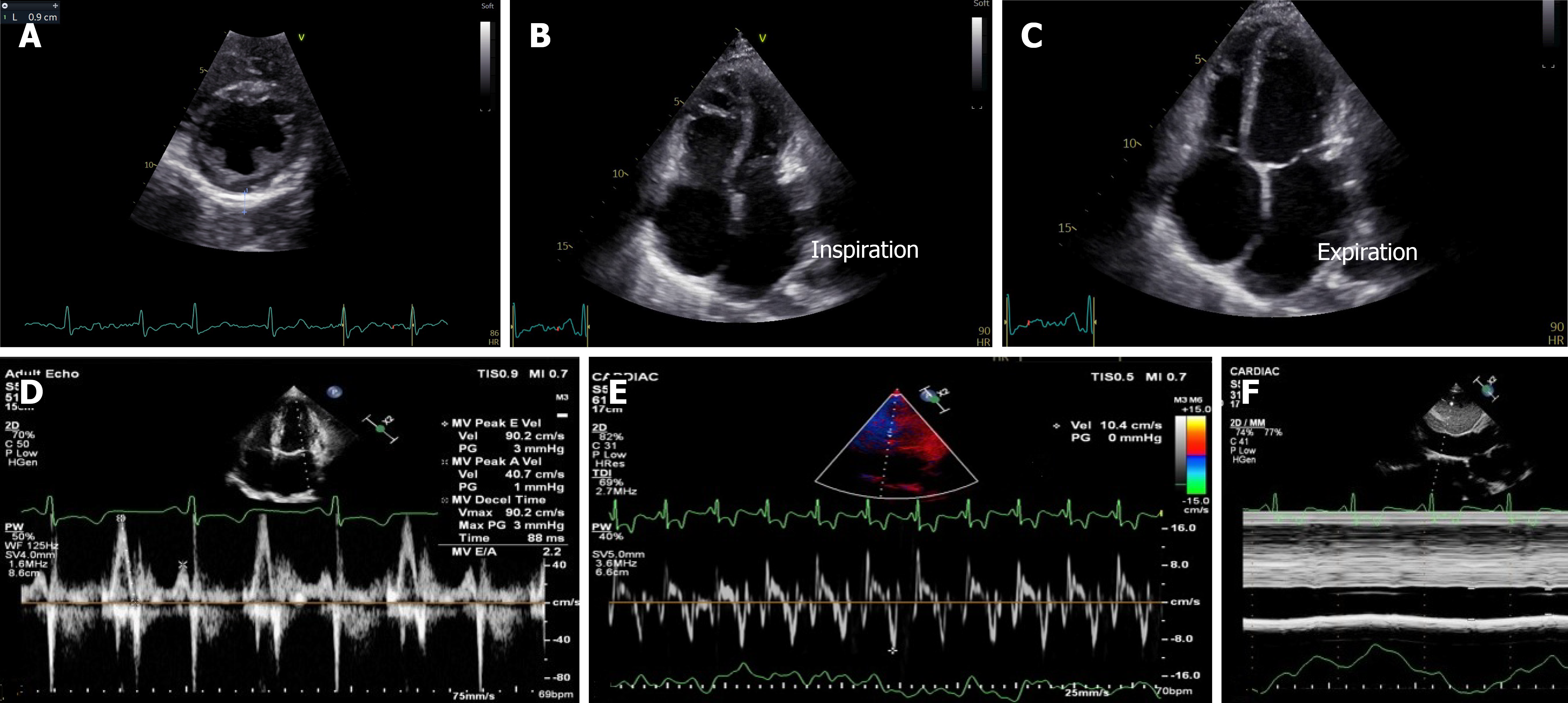
Figure 4 Transthoracic echocardiography.
A: Thickened and calcified pericardium in left parasternal short-axis view; B and C: Respirophasic shift of interventricular septum in apical 4-chamber view; D: Mitral valve E/A 2.2 in pulsed wave Doppler echocardiography; E: Medial mitral annular velocity 10.4 cm/second in tissue Doppler imaging; F: Dilated, non-collapsing inferior vena cava in two-dimensional-guided M-mode echocardiography in subcostal view.
- Citation: Islam AKMM. Constrictive pericarditis in the twenty-first century: An old disease revisited. World J Cardiol 2026; 18(8): 121750
- URL: https://www.wjgnet.com/1949-8462/full/v18/i8/121750.htm
- DOI: https://dx.doi.org/10.4330/wjc.121750