
















Copyright: ©Author(s) 2026.
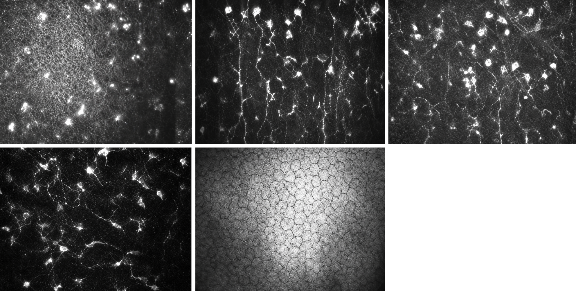
Figure 3 In vivo confocal microscopy findings in amiodarone-related keratopathy.
Representative confocal images show numerous hyperreflective intracellular inclusions predominantly within the corneal epithelial layers, consistent with drug-induced phospholipid deposition. These images illustrate the microstructural substrate of cornea verticillata and support the role of confocal microscopy as an adjunctive tool for documenting epithelial involvement in vivo.
- Citation: Capobianco M, Nicolosi SG, Cappellani F, Avitabile A, Cannizaro L, Visalli F, D’Esposito F, Gattazzo I, Gagliano C, Zeppieri M. Identifying and treating amiodarone-induced corneal deposits: When the heart saves and the cornea speaks. World J Cardiol 2026; 18(6): 121185
- URL: https://www.wjgnet.com/1949-8462/full/v18/i6/121185.htm
- DOI: https://dx.doi.org/10.4330/wjc.121185