
















Copyright: ©Author(s) 2026.
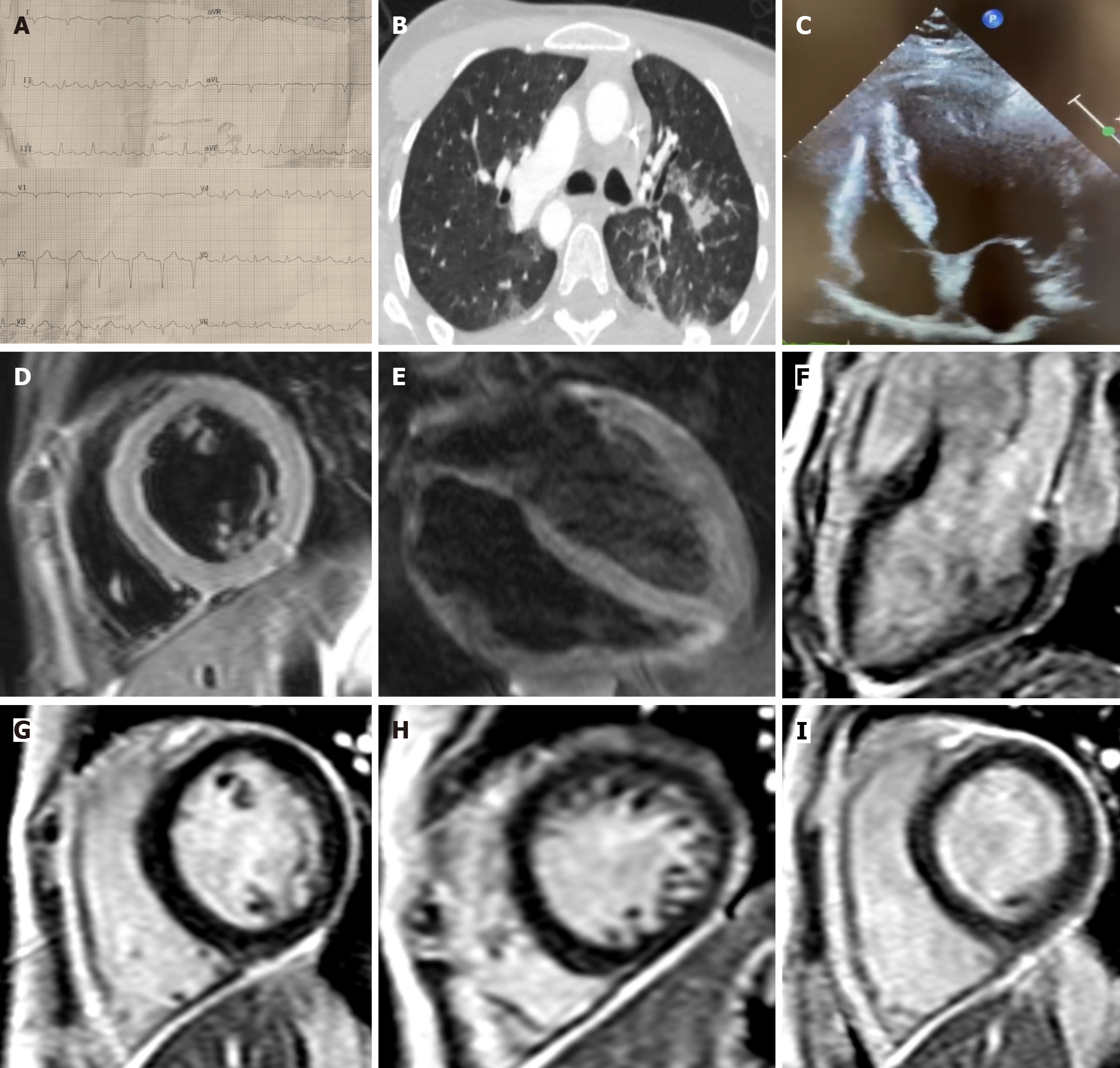
Figure 1 Multimodal assessment of the patient during coronavirus disease 2019 related myocarditis.
A: The 12-lead electrocardiogram at admission revealed sinus tachycardia and ST-segment elevation in the inferolateral leads, without reciprocal ST-segment depression in the corresponding leads; B: Chest computed tomography scan performed during coronavirus disease 2019 infection, revealing bilateral ground-glass opacities and consolidations; C: Echocardiographic apical four chambers view during cardiogenic shock; D-I: Cardiac magnetic resonance revealed an area of edema in the mid-basal inferior, inferolateral and lateral walls and a non-ischemic late gadolinium enhancement pattern with thin meso-/subepicardial striae in the inferior and inferolateral walls of left ventricle.
- Citation: Birtolo LI, Ferranti F, Manzi G, Caputo A, Scoccia G, Bruno K, Galea N, Maestrini V, Chimenti C, Severino P, Pugliese F, Badagliacca R, Vizza CD. From COVID-19 to influenza A - recurrent viral myocarditis and successful immunosuppressive therapy: A case report and review of literature. World J Cardiol 2026; 18(6): 113066
- URL: https://www.wjgnet.com/1949-8462/full/v18/i6/113066.htm
- DOI: https://dx.doi.org/10.4330/wjc.113066