
















Copyright: ©Author(s) 2026.
World J Cardiol. Apr 26, 2026; 18(4): 118863
Published online Apr 26, 2026. doi: 10.4330/wjc.v18.i4.118863
Published online Apr 26, 2026. doi: 10.4330/wjc.v18.i4.118863
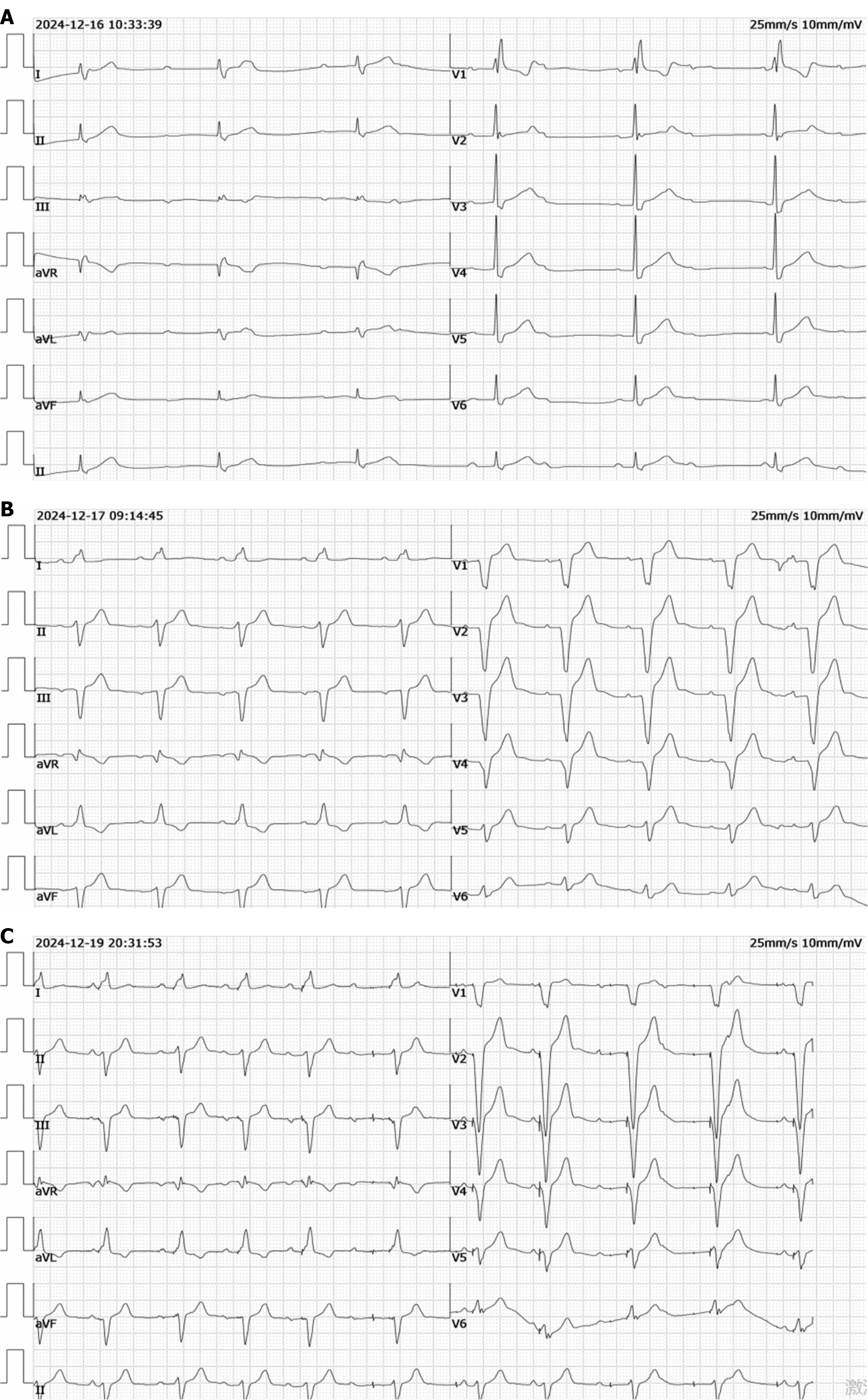
Figure 1 Changes in the electrocardiogram before and after pacemaker implantation.
A: Upon admission, the patient’s electrocardiogram (ECG) showed third-degree atrioventricular block; B: At the end of pacemaker implantation, the ECG showed a pacemaker operating mode of ventricular pacing with atrial sensing and ventricular triggering, indicating normal atrial sensing and ventricular pacing functions; C: On the third postoperative day, when diaphragmatic flutter occurred, the ECG showed a dual-chamber pacing, dual-chamber sensing, dual-response mode. Atrial sensing and pacing were impaired, whereas ventricular sensing and pacing remained normal. The QRS complexes of the 2nd-5th ventricular paced beats in the limb leads were all triggered by previously sensed intrinsic atrial waves. An inappropriate atrial pulse was delivered before the QRS complex of the 6th ventricular paced beat, suggesting abnormal sensing. No atrial wave followed the pulse, suggesting abnormal pacing.
- Citation: Zhang XS, Wang F, Qi XW, Shafii P, Cui XN, Wang YH. Phrenic nerve capture caused by atrial electrode dislodgement: A case report. World J Cardiol 2026; 18(4): 118863
- URL: https://www.wjgnet.com/1949-8462/full/v18/i4/118863.htm
- DOI: https://dx.doi.org/10.4330/wjc.v18.i4.118863