
















Copyright: ©Author(s) 2026.
World J Cardiol. Apr 26, 2026; 18(4): 118546
Published online Apr 26, 2026. doi: 10.4330/wjc.v18.i4.118546
Published online Apr 26, 2026. doi: 10.4330/wjc.v18.i4.118546
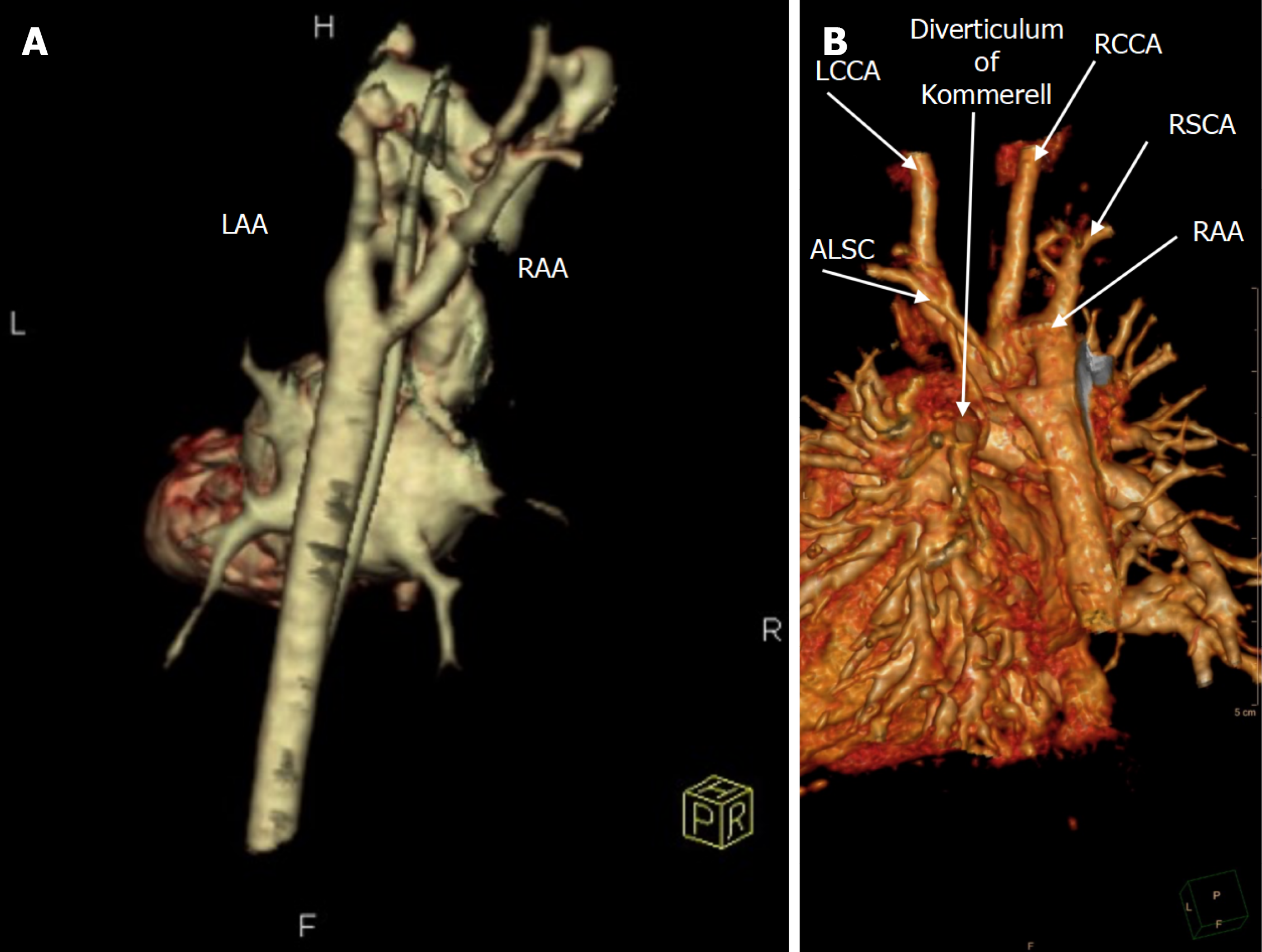
Figure 4 Three-dimensional computed tomography angiography of patient.
A: Three-dimensional computed tomography angiography (CTA) reconstruction of patient 5 demonstrating the vascular anatomy in a patient with double aortic arch, showing both arches encircling the trachea and esophagus. Double aortic arch is the most common complete vascular ring; identifying the dominant vs nondominant arch on CTA is essential because it determines which arch segment is divided during surgical repair; B: Three-dimensional volume-rendered CTA demonstrating a right aortic arch with an aberrant left subclavian artery (ALSA) arising from a Kommerell diverticulum, with labeled branch vessels and the diverticulum origin of the ALSA. In a right aortic arch, identifying an ALSA arising from a Kommerell diverticulum is crucial because the diverticulum and retroesophageal ALSA can be the dominant compressive components; recognizing this anatomy preoperatively guides whether simple ligamentum division is sufficient or whether diverticulum/subclavian-directed intervention may be needed to prevent persistent symptoms. LAA: Left aortic arch; RAA: Right aortic arch; LCCA: Left common carotid artery; RCCA: Right common carotid artery; RSCA: Right subclavian artery; ALSC: Aberrant left subclavian.
- Citation: Abdul Khalek J, Kanbar K, Zareef R, Alzein MH, El-Rassi I, Bitar FF, Arabi MT. Vascular rings in a tertiary care center: A retrospective review of clinical presentations and interventions. World J Cardiol 2026; 18(4): 118546
- URL: https://www.wjgnet.com/1949-8462/full/v18/i4/118546.htm
- DOI: https://dx.doi.org/10.4330/wjc.v18.i4.118546