
















Copyright: ©Author(s) 2026.
World J Cardiol. Mar 26, 2026; 18(3): 117999
Published online Mar 26, 2026. doi: 10.4330/wjc.v18.i3.117999
Published online Mar 26, 2026. doi: 10.4330/wjc.v18.i3.117999
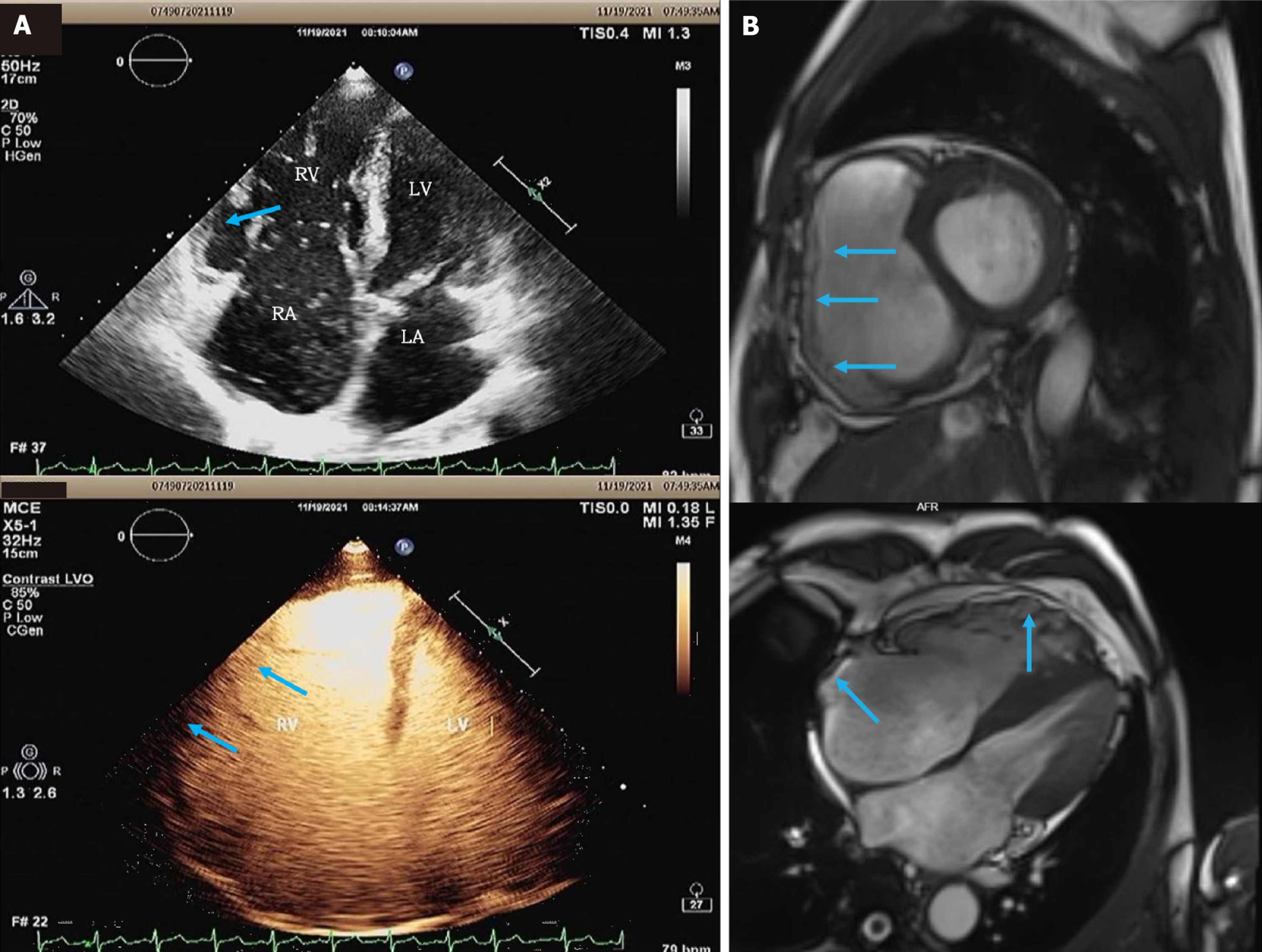
Figure 3 Results of echocardiography and cardiac magnetic resonance.
A: Contrast echocardiography showing no significant abnormalities in left ventricular wall motion, right ventricle (RV) wall motion abnormalities, RV fractional area change measured at approximately 30%, RV aneurysm (blue arrows); B: Cardiac magnetic resonance showing fatty infiltration of the RV free wall and the RA (blue arrows).
- Citation: Li A, Zhu YJ, Ma XX, Jin Y, Yang J, Feng QT. Arrhythmogenic right ventricular cardiomyopathy with atrial tachycardia and sick sinus syndrome: A case report. World J Cardiol 2026; 18(3): 117999
- URL: https://www.wjgnet.com/1949-8462/full/v18/i3/117999.htm
- DOI: https://dx.doi.org/10.4330/wjc.v18.i3.117999