











BPG is committed to discovery and dissemination of knowledge
Basic Study
Copyright: ©Author(s) 2026.
World J Cardiol. Mar 26, 2026; 18(3): 117821
Published online Mar 26, 2026. doi: 10.4330/wjc.v18.i3.117821
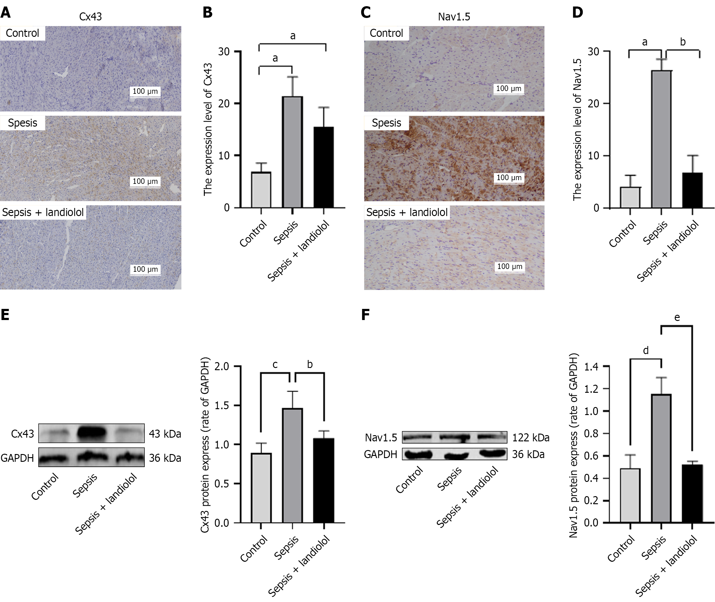
Figure 6
Figure 6 Comparative analysis of connexin-43 expression via immunohistochemistry across the experimental groups. A: Immunohistochemical staining results of connexin-43 (Cx43) channel protein in ventricular myocardial tissues of rats; B: Quantitative analysis of Cx43 expression levels in ventricular myocardial tissues; C: Immunohistochemical staining results of sodium 1.5 ion (Nav1.5) channel protein in ventricular myocardial tissues of rats; D: Quantitative analysis of Nav1.5 channel expression levels in ventricular myocardial tissues; E and F: Protein expression levels of Cx43/Nav1.5 channel in rat ventricular myocardial tissue were detected by western blot, with glyceraldehyde-3-phosphate dehydrogenase as the internal control. Quantitative analysis of the above protein expressions was conducted. aP < 0.05 vs control, bP < 0.05 vs sepsis group, cP < 0.01 vs control, dP < 0.001 vs control, eP < 0.001 vs sepsis group and n = 3. Original magnification was 20 ×, n = 3. All data are expressed as mean ± SD. Cx43: Connexin-43; Nav1.5: Sodium 1.5 ion; GAPDH: Glyceraldehyde-3-phosphate dehydrogenase.


Write to the Help Desk