











BPG is committed to discovery and dissemination of knowledge
Basic Study
Copyright: ©Author(s) 2026.
World J Cardiol. Mar 26, 2026; 18(3): 117821
Published online Mar 26, 2026. doi: 10.4330/wjc.v18.i3.117821
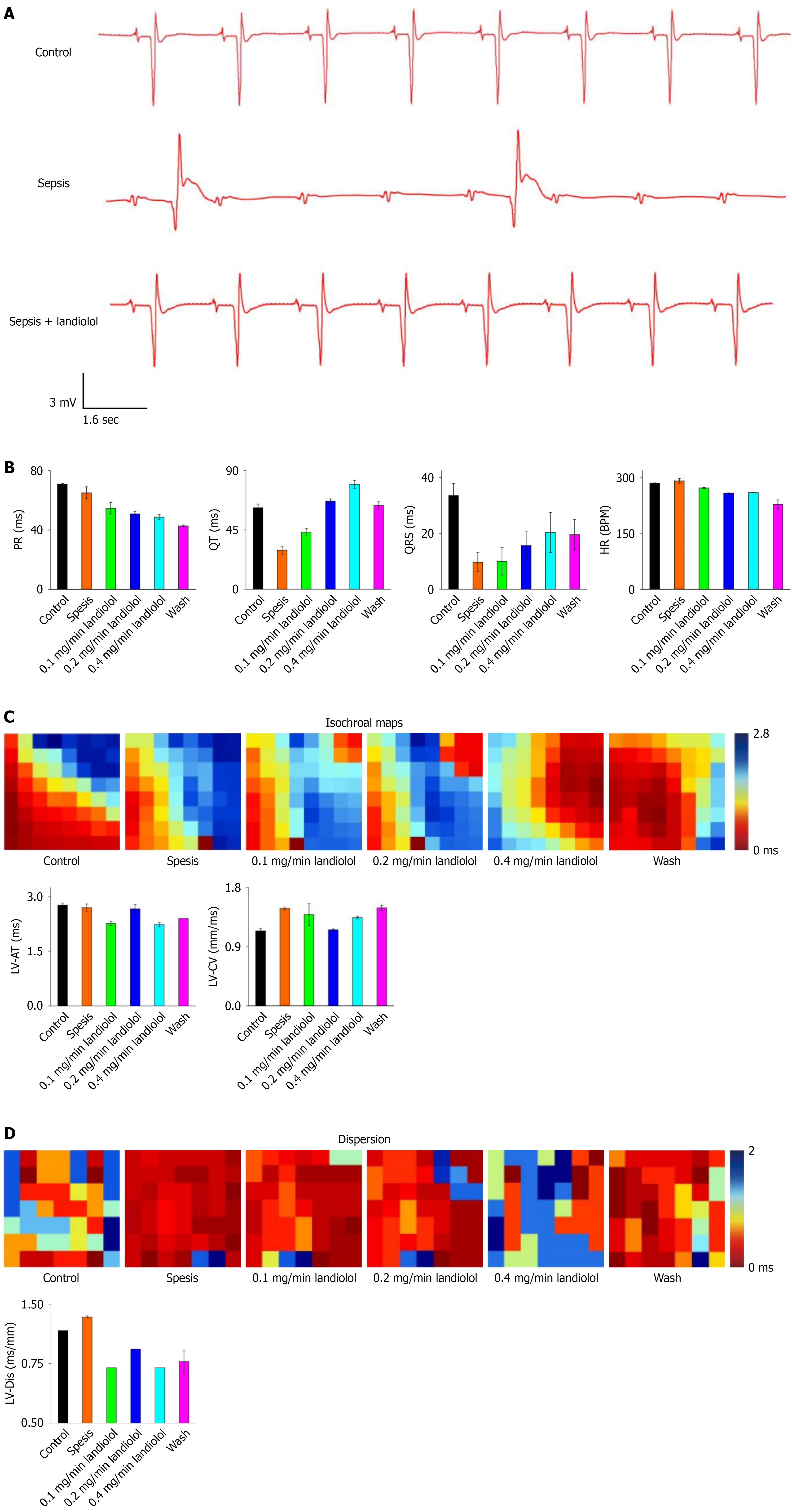
Figure 5
Figure 5 Landiolol reverses the abnormal conduction velocity of ventricular myocytes in septic rats, and significantly reduces conduction dispersion and heart rate. A: Electrocardiographic recordings from isolated perfused hearts of rats in each experimental group; B: Comparison of various interval durations (PR, QT, QTS) and heart rates in isolated perfused hearts among the three groups; C: Comparative analysis of conduction time and conduction velocity in isolated perfused hearts in the experimental groups, left ventricular active time, left ventricular contraction velocity; D: Comparative analysis of conduction dispersion in isolated perfused hearts across the experimental groups. Black represents the control group, red represents the sepsis model group, green represents the sepsis model group treated with low-dose (0.1 mg/minute) landiolol, dark blue represents the sepsis model group treated with medium-dose (0.2 mg/minute) landiolol, light blue represents the sepsis model group treated with high-dose (0.4 mg/minute) landiolol, and pink represents the landiolol washout group. n = 3. HR: Heart rates; LV-AT: Left ventricular active time; LV-CV: Left ventricular contraction velocity.


Write to the Help Desk