
















Copyright: ©Author(s) 2026.
World J Cardiol. Mar 26, 2026; 18(3): 116299
Published online Mar 26, 2026. doi: 10.4330/wjc.v18.i3.116299
Published online Mar 26, 2026. doi: 10.4330/wjc.v18.i3.116299
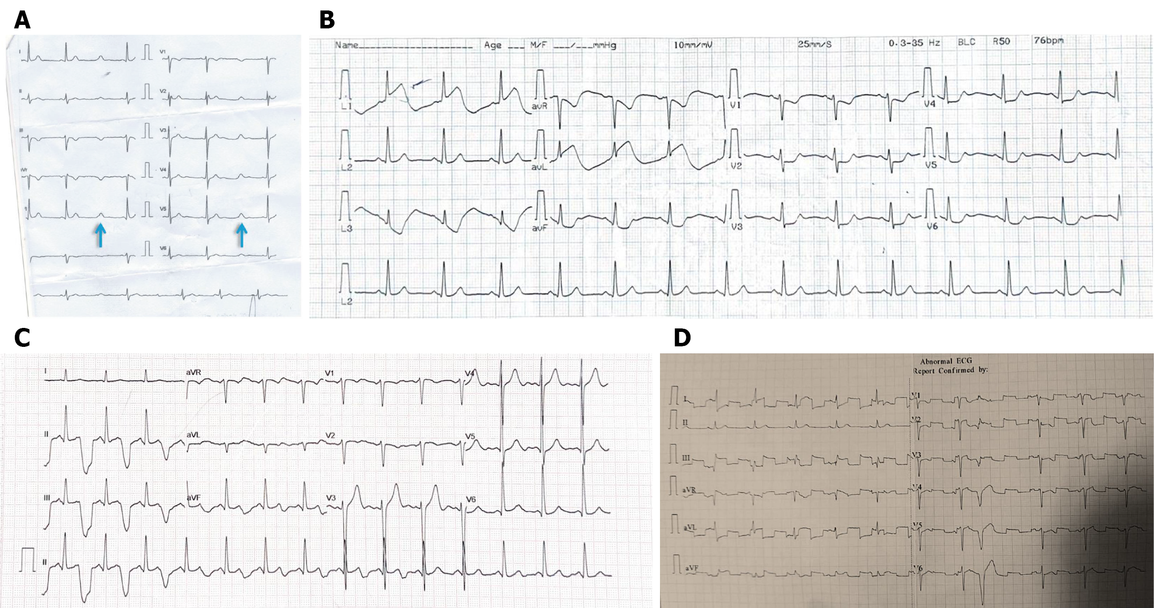
Figure 7 Equipment-related and environmental artifacts.
A: Missing PQRSs due to processing failure with residual T waves give rise to double T waves or “orphan T” waves (arrows); B: Arterial pulse tapping artifact mimicking ST elevation. All QRS complexes are followed by artifacts with electromechanical associations. Note that lead 2 is spared, meaning that the artifact comes from the left arm electrode; C: Similarly, arterial pulse tapping artifacts mimic bizarre T waves with prolonged QT intervals. Note that lead 1 is spared, meaning that the artifact comes from the left lower limb electrode; D: Electrocardiogram showing fixed-frequency square wave artifacts. ECG: Electrocardiogram.
- Citation: Mondal S, Raja DP, Muslim NA, Prabhu MA. Electrocardiographic artifacts in clinical practice: A logical approach to recognition and prevention. World J Cardiol 2026; 18(3): 116299
- URL: https://www.wjgnet.com/1949-8462/full/v18/i3/116299.htm
- DOI: https://dx.doi.org/10.4330/wjc.v18.i3.116299