
















Copyright: ©Author(s) 2026.
World J Cardiol. Mar 26, 2026; 18(3): 116299
Published online Mar 26, 2026. doi: 10.4330/wjc.v18.i3.116299
Published online Mar 26, 2026. doi: 10.4330/wjc.v18.i3.116299
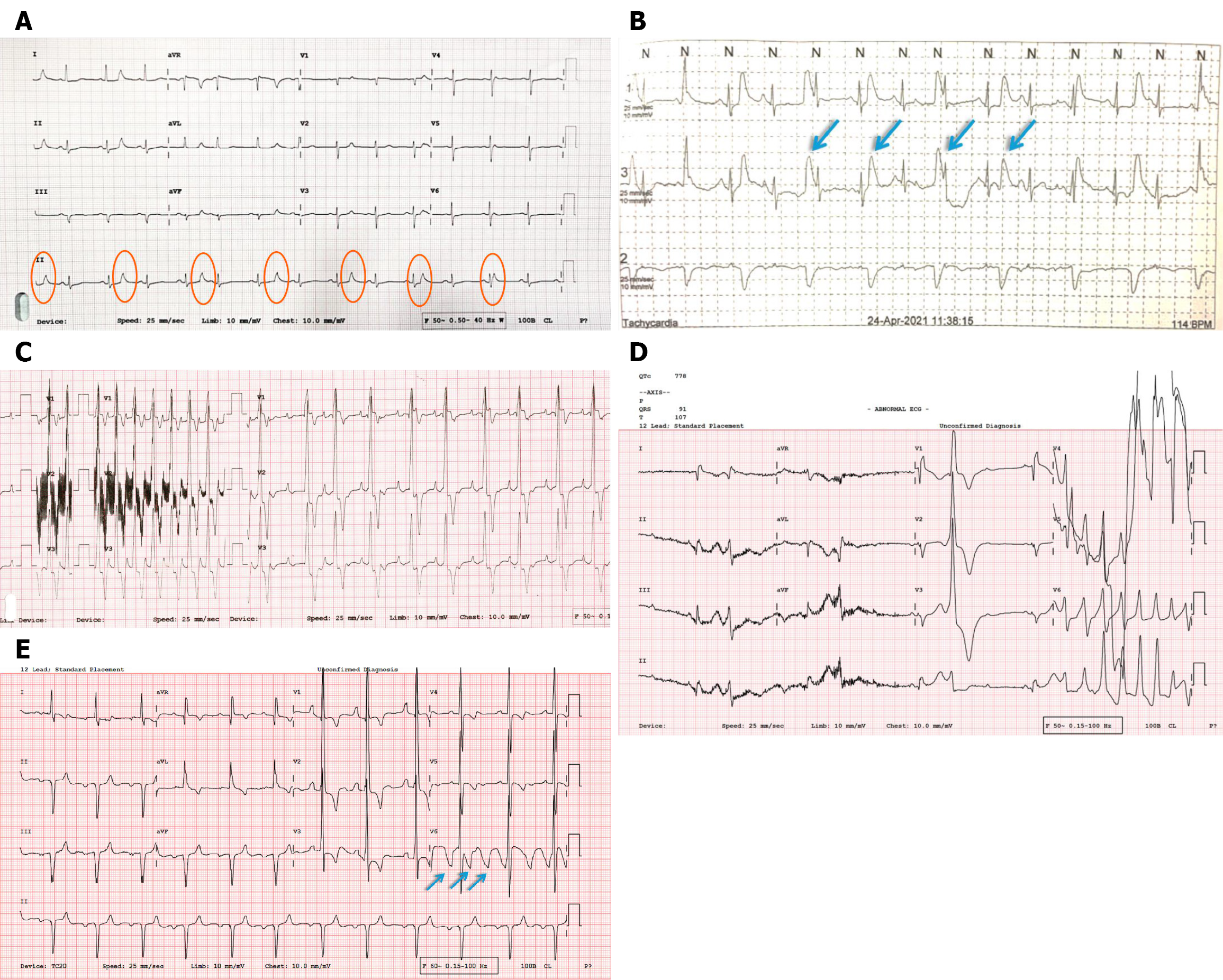
Figure 5 Patient-related and equipment-related artifacts.
A: Low-frequency artifacts (0.7 Hz) can be seen marching through without any PQRST association (orange circles). While the source of the artifact is unclear, they are too slow to be physiological tremors, which fall in the range of 3-12 Hz (or even higher). Respiratory movements can typically cause artifacts in this range (0.7 Hz), but the odd point corresponds to a respiratory rate of approximately 42/minute. The heart rate is too slow for that type of tachypnea; B: Similar artifacts can be seen on a Holter tracing from a young child, likely a respiration artifact (arrows); C: Attenuation correction artifacts showing low-amplitude high-frequency noise picked up by V2 lead to a crying newborn (D/D intercostal muscle fasciculation); D: Poor electrode contact causes intermittent baseline jumps in V4 and V5. Notably, artifacts may coexist with life-threatening arrhythmias. In this example, the patient had bradycardia-related Torsades de Pointes with simultaneous artifacts, emphasizing clinical evaluation while interpreting or suspecting artifacts on electrocardiogram; E: Poor electrode contact causes irregular baseline jumps in V6 (arrows).
- Citation: Mondal S, Raja DP, Muslim NA, Prabhu MA. Electrocardiographic artifacts in clinical practice: A logical approach to recognition and prevention. World J Cardiol 2026; 18(3): 116299
- URL: https://www.wjgnet.com/1949-8462/full/v18/i3/116299.htm
- DOI: https://dx.doi.org/10.4330/wjc.v18.i3.116299