
















Copyright: ©Author(s) 2026.
World J Cardiol. Mar 26, 2026; 18(3): 116299
Published online Mar 26, 2026. doi: 10.4330/wjc.v18.i3.116299
Published online Mar 26, 2026. doi: 10.4330/wjc.v18.i3.116299
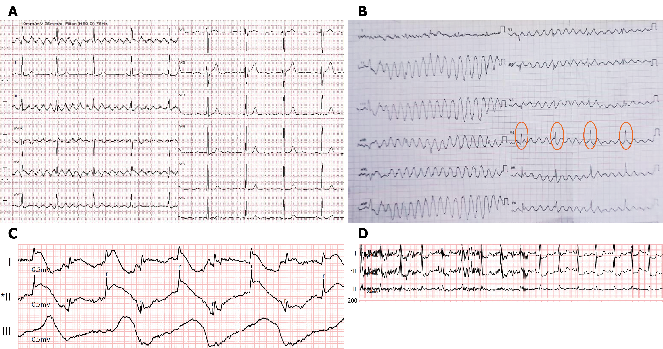
Figure 4 Patient-related motion artifacts.
A: Classical Parkinson’s rest tremor. Note that the frequency of the artifact is approximately 5 Hz (200 microseconds interval). Note that lead 2 does not have an artifact, indicating that the artifact is coming from the left arm; B: Limb movement artifacts resembling polymorphic ventricular tachycardia. Note that the normal QRS complexes (orange circles) are marching through the artifacts; C: Intraprocedural spurious ST elevation due to lead movement (attached to the subcostal region) during respiration; D: Trembling during micturition, causing baseline high-frequency irregular artifacts; note that the stoppage of the act of micturition resolves the baseline artifact.
- Citation: Mondal S, Raja DP, Muslim NA, Prabhu MA. Electrocardiographic artifacts in clinical practice: A logical approach to recognition and prevention. World J Cardiol 2026; 18(3): 116299
- URL: https://www.wjgnet.com/1949-8462/full/v18/i3/116299.htm
- DOI: https://dx.doi.org/10.4330/wjc.v18.i3.116299