
















Copyright: ©Author(s) 2026.
World J Cardiol. Mar 26, 2026; 18(3): 116115
Published online Mar 26, 2026. doi: 10.4330/wjc.v18.i3.116115
Published online Mar 26, 2026. doi: 10.4330/wjc.v18.i3.116115
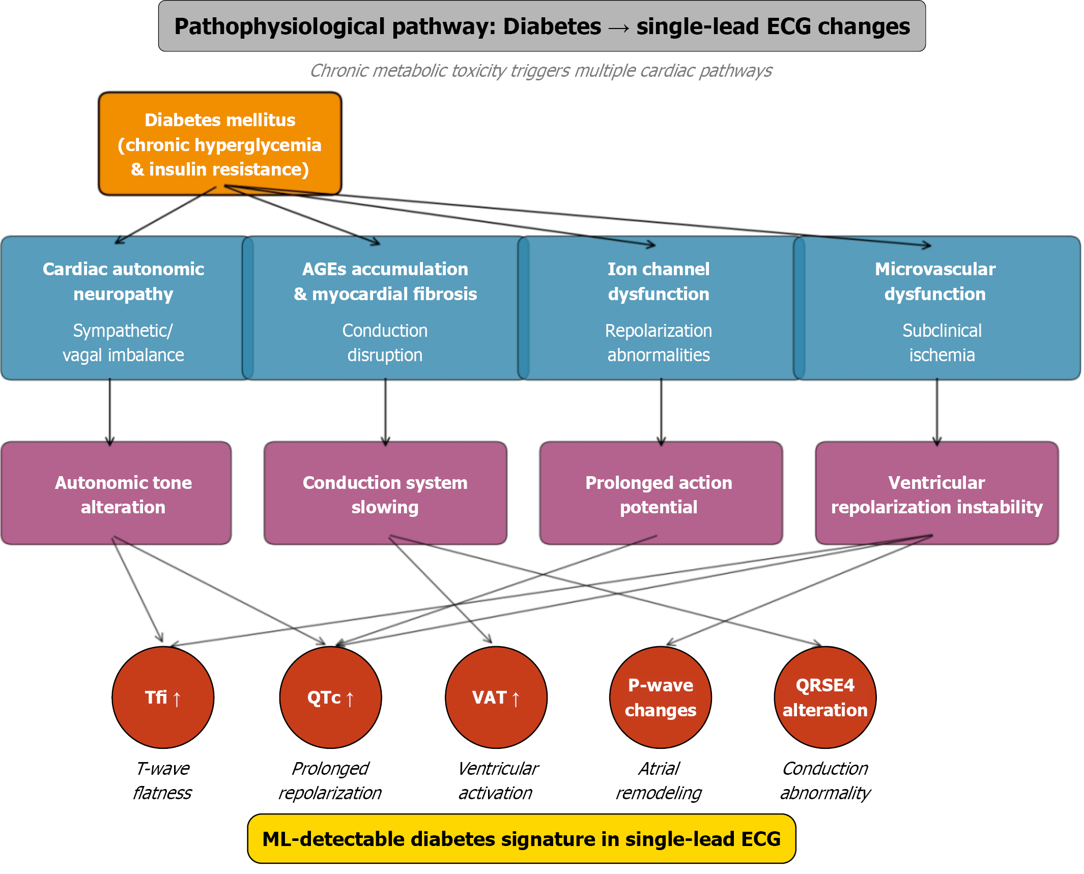
Figure 4 Pathophysiological pathway of diabetes-induced cardiac electrophysiological changes detected by single-lead electrocar
- Citation: Karbovskaya AD, Marzoog BA, Stroeva A, Chomakhidze P, Gognieva D, Kuznetsova N, Syrkin A, Fadeev VV, Poluboyarinova IV, Ismailova SM, Suvorov A, Kopylov P. Discriminating diabetes mellitus from single-lead electrocardiography using machine learning and multinomial regression. World J Cardiol 2026; 18(3): 116115
- URL: https://www.wjgnet.com/1949-8462/full/v18/i3/116115.htm
- DOI: https://dx.doi.org/10.4330/wjc.v18.i3.116115