
















©The Author(s) 2026.
World J Cardiol. Feb 26, 2026; 18(2): 113358
Published online Feb 26, 2026. doi: 10.4330/wjc.v18.i2.113358
Published online Feb 26, 2026. doi: 10.4330/wjc.v18.i2.113358
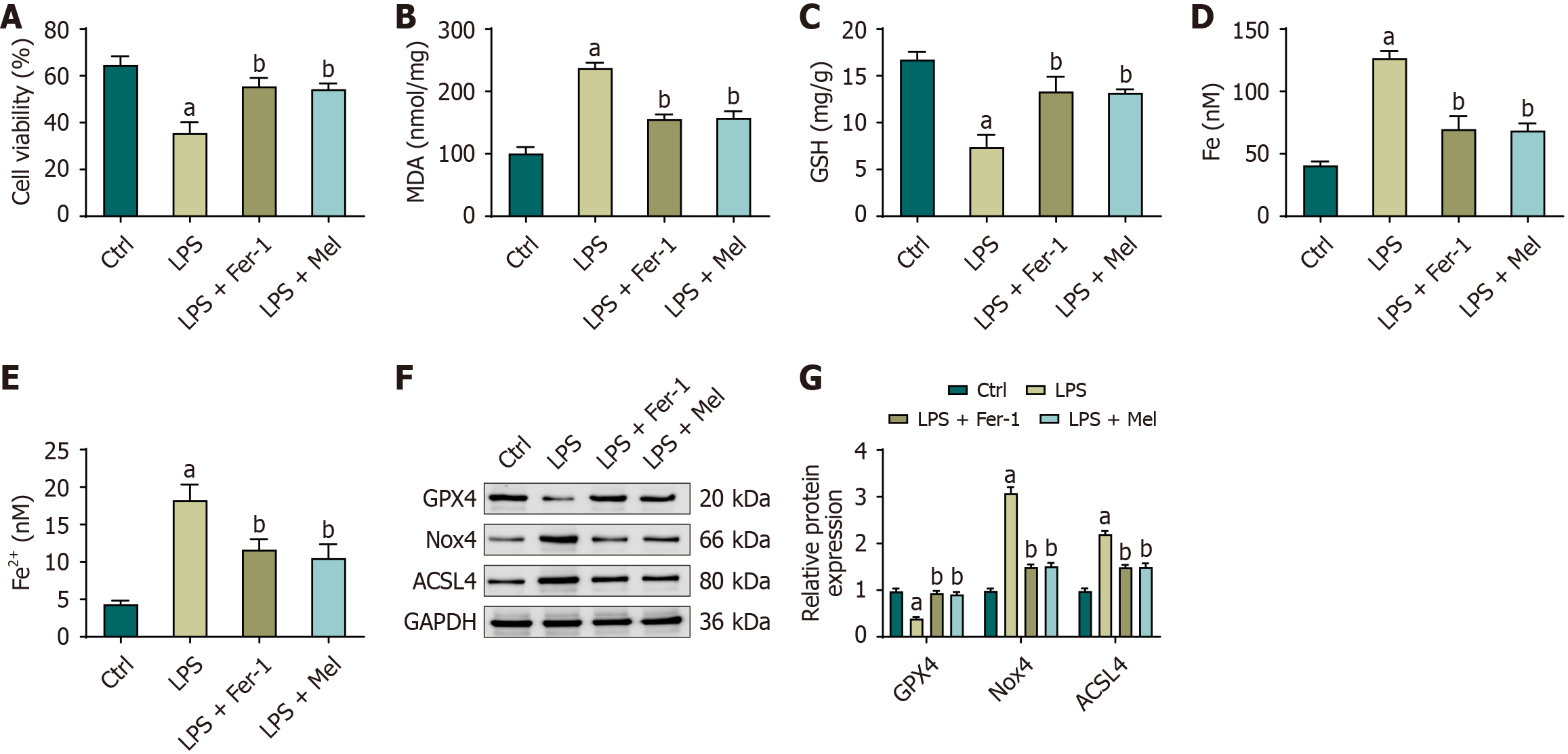
Figure 4 Melatonin improves viability, redox balance, and ferroptosis markers in HL-1 cardiomyocytes.
A: Cell viability assessed by cell counting kit-8; B-E: Cellular malondialdehyde, glutathione, total iron, and ferrous iron after treatment with lipopolysaccharide with or without ferrostatin-1 or melatonin; F: Representative immunoblots of glutathione peroxidase 4, NADPH oxidase 4, and acyl-CoA synthetase long-chain family member 4 (glyceraldehyde-3-phosphate dehydrogenase as control); G: Densitometric quantification normalized to glyceraldehyde-3-phosphate dehydrogenase. Data are presented as mean ± SD from three independent biological experiments (each measured in technical triplicate). One-way analysis of variance with Bonferroni post-hoc testing. aP < 0.01 vs control; bP < 0.01 vs lipopolysaccharide. ACSL4: Acyl-CoA synthetase long-chain family member 4; Ctrl: Control; Fe2+: Ferrous iron; GAPDH: Glyceraldehyde-3-phosphate dehydrogenase; GPX4: Glutathione peroxidase 4; GSH: Glutathione; LPS: Lipopolysaccharide; LPS + Mel: Lipopolysaccharide plus melatonin; LPS + Fer-1: Lipopolysaccharide plus ferrostatin-1; MDA: Malondialdehyde; Nox4: NADPH oxidase 4.
- Citation: Zeng M, Li Q, Li L, Xiang CF, Wang YJ. Melatonin regulates Sirt1/Nrf2/GPX4 pathway to inhibit ferroptosis and alleviate myocardial injury caused by sepsis. World J Cardiol 2026; 18(2): 113358
- URL: https://www.wjgnet.com/1949-8462/full/v18/i2/113358.htm
- DOI: https://dx.doi.org/10.4330/wjc.v18.i2.113358