
















©The Author(s) 2026.
World J Cardiol. Jan 26, 2026; 18(1): 112857
Published online Jan 26, 2026. doi: 10.4330/wjc.v18.i1.112857
Published online Jan 26, 2026. doi: 10.4330/wjc.v18.i1.112857
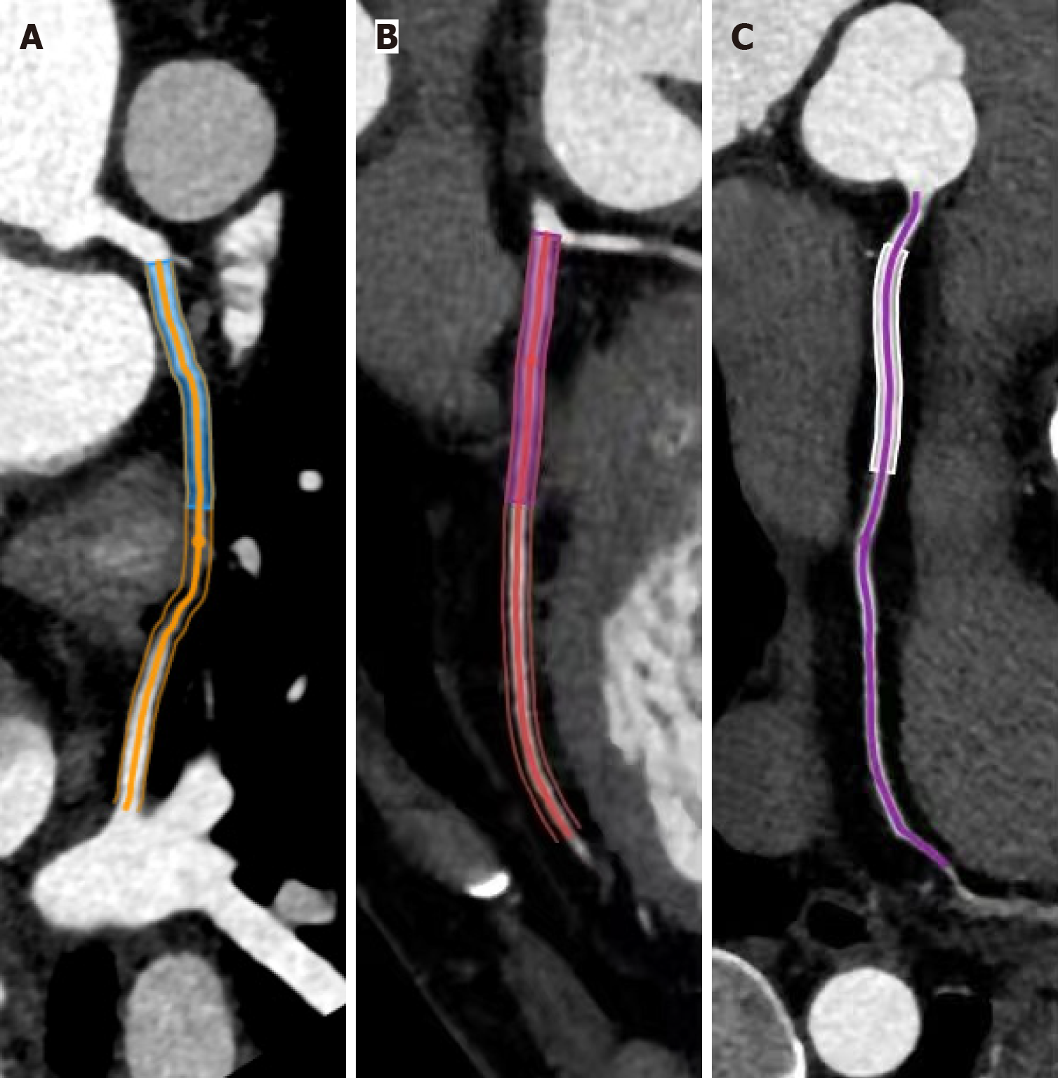
Figure 1 The methodology of the pericoronary adipose tissue analysis.
A: Left anterior descending artery; B: Left circumflex artery proximal 40-mm segments; C: Right coronary artery proximal 10-50 mm segment. Pericoronary adipose tissue attenuation was quantified within the adipose voxels (-190 HU to -30 HU), extending radially from the vessel wall at distances equivalent to the coronary artery diameters.
- Citation: Su KX, Jiang SY, Pang CF, Yang F, Tang YQ, Li XG, He WF, Li R. Coronary computed tomography angiography adipose tissue attenuation in diabetic patients with myocardial ischemia and non-obstructive coronary arteries. World J Cardiol 2026; 18(1): 112857
- URL: https://www.wjgnet.com/1949-8462/full/v18/i1/112857.htm
- DOI: https://dx.doi.org/10.4330/wjc.v18.i1.112857