
















©The Author(s) 2026.
World J Cardiol. Jan 26, 2026; 18(1): 112636
Published online Jan 26, 2026. doi: 10.4330/wjc.v18.i1.112636
Published online Jan 26, 2026. doi: 10.4330/wjc.v18.i1.112636
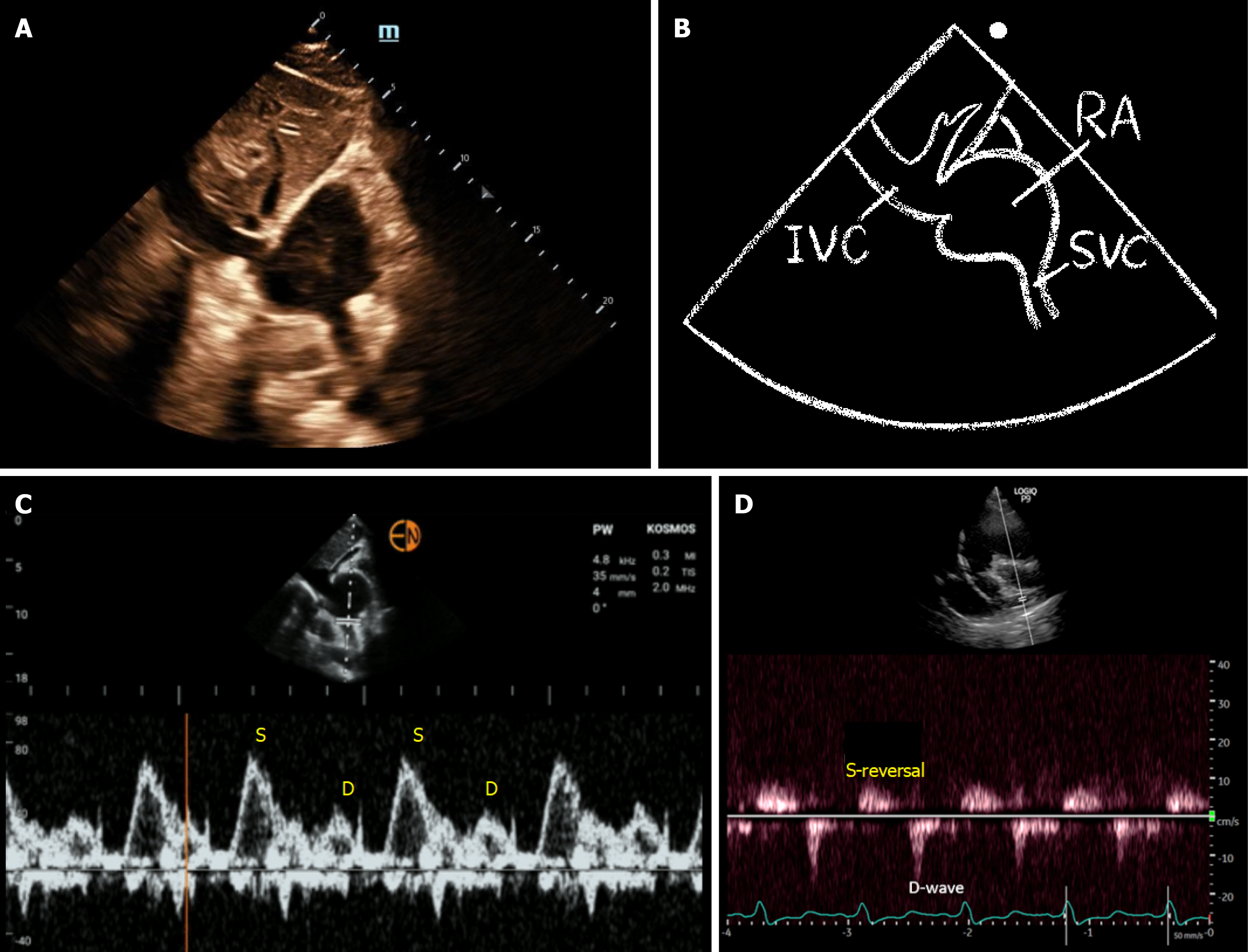
Figure 4 Superior vena cava Doppler.
A and B: Subcostal bicaval view with corresponding illustration; C and D: Panel C shows a normal superior vena cava Doppler waveform obtained from the bicaval view with a systolic wave greater than the diastolic wave. Panel D demonstrates a severely abnormal pattern with systolic wave reversal. The tracing in panel D was obtained from a modified parasternal view, which often results in suboptimal Doppler angle and consequently lower recorded flow velocities. IVC: Inferior vena cava; RA: Right atrium; SVC: Superior vena cava; S: Systolic; D: Diastolic.
- Citation: Koratala A. Extended venous excess ultrasound: A promising addition to point-of-care ultrasound-based venous congestion assessment. World J Cardiol 2026; 18(1): 112636
- URL: https://www.wjgnet.com/1949-8462/full/v18/i1/112636.htm
- DOI: https://dx.doi.org/10.4330/wjc.v18.i1.112636