Copyright
©The Author(s) 2025.
World J Cardiol. Jul 26, 2025; 17(7): 108745
Published online Jul 26, 2025. doi: 10.4330/wjc.v17.i7.108745
Published online Jul 26, 2025. doi: 10.4330/wjc.v17.i7.108745
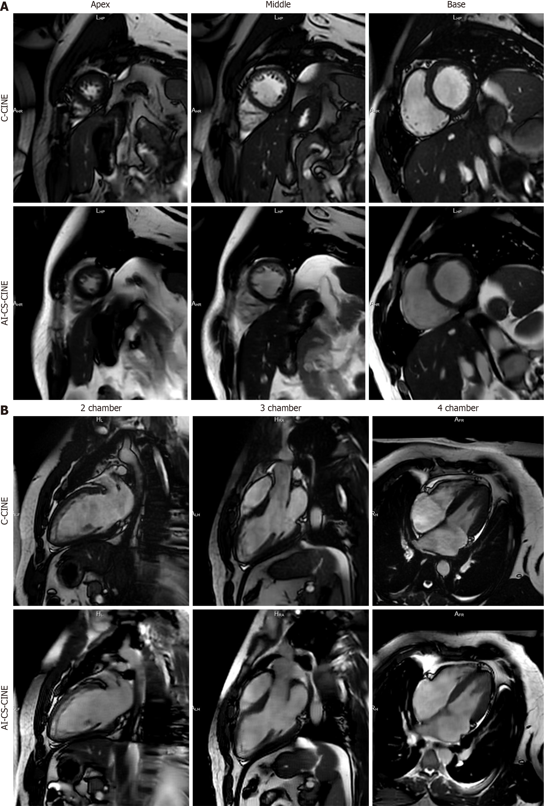
Figure 2 Magnetic resonance imaging illustrating the exceptional alignment between conventional CINE and artificial-intelligence-assisted compressed sensing CINE in a healthy 20-year-old male volunteer.
A: The short-axis stack clearly depicts the apex, middle, and base of the left ventricle in both conventional CINE (C-CINE) (top row) and artificial-intelligence-assisted compressed sensing CINE (AI-CS-CINE) (bottom row); B: Additionally, the 2-, 3-, and 4-chamber views demonstrate excellent agreement between C-CINE and AI-CS-CINE in this volunteer. C-CINE: Conventional CINE; AI-CS-CINE: Artificial-intelligence-assisted compressed sensing CINE.
- Citation: Wang H, Schmieder A, Watkins M, Wang P, Mitchell J, Qamer SZ, Lanza G. Artificial intelligence-assisted compressed sensing CINE enhances the workflow of cardiac magnetic resonance in challenging patients. World J Cardiol 2025; 17(7): 108745
- URL: https://www.wjgnet.com/1949-8462/full/v17/i7/108745.htm
- DOI: https://dx.doi.org/10.4330/wjc.v17.i7.108745


