Copyright
©The Author(s) 2025.
World J Cardiol. Jun 26, 2025; 17(6): 107729
Published online Jun 26, 2025. doi: 10.4330/wjc.v17.i6.107729
Published online Jun 26, 2025. doi: 10.4330/wjc.v17.i6.107729
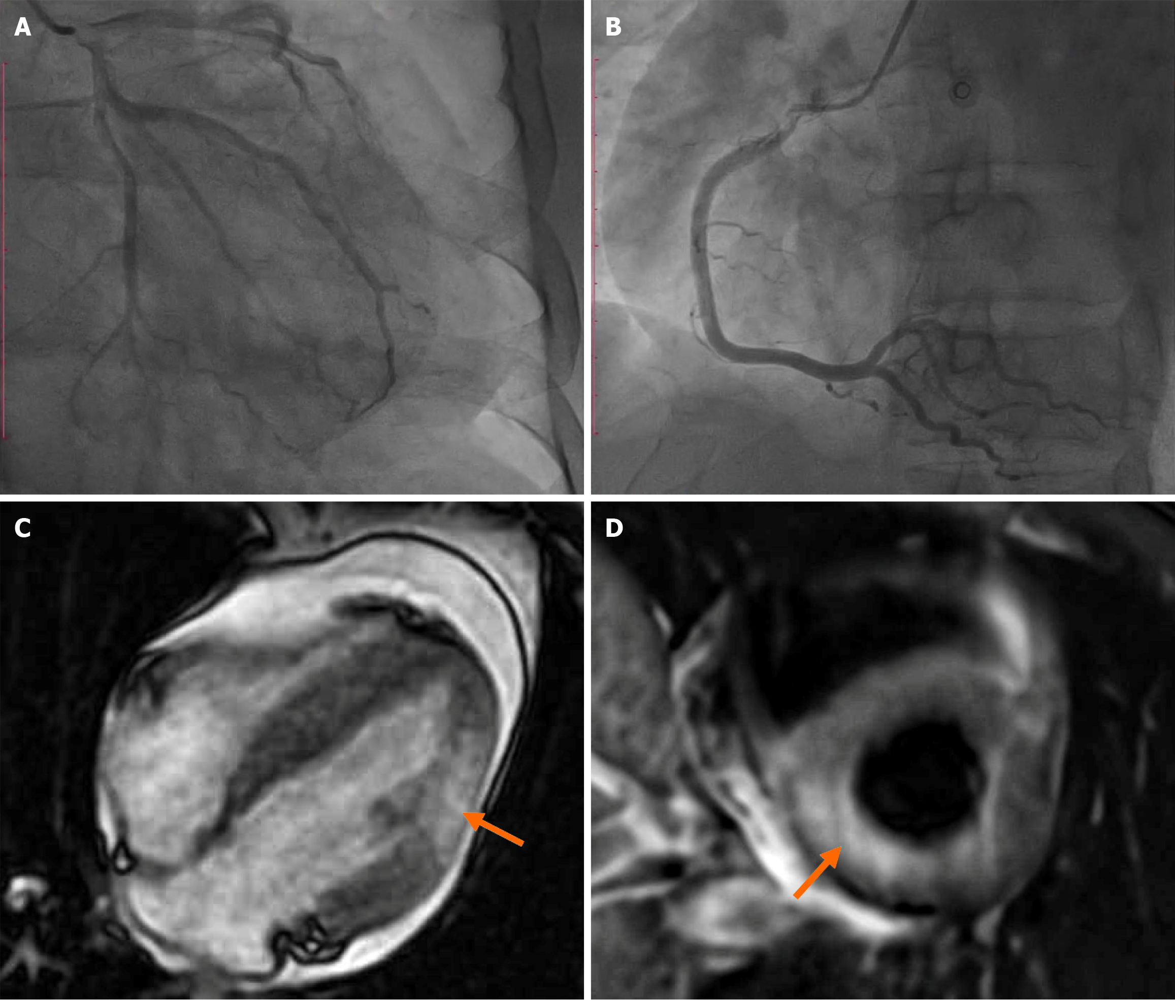
Figure 1 Coronary angiography and cardiac magnetic resonance imaging findings in a patient with suspected myocarditis.
A and B: Coronary angiography reveals normal coronary anatomy with no evidence of luminal stenosis, effectively excluding obstructive coronary artery disease; C and D: Cardiac magnetic resonance imaging demonstrates focal myocardial injury indicated by arrows, characterized by increased signal intensity on T2-weighted imaging (edema) and late gadolinium enhancement in a subepicardial distribution of the lateral wall, consistent with acute myocarditis.
- Citation: Luong TV, Hoang TA, Pham NN, Nguyen STM, Tran QB, Nguyen HM, Doan TC, Ho BA, Dang HNN. Eosinophilic myocarditis due to parasitic infection: A case-based minireview. World J Cardiol 2025; 17(6): 107729
- URL: https://www.wjgnet.com/1949-8462/full/v17/i6/107729.htm
- DOI: https://dx.doi.org/10.4330/wjc.v17.i6.107729


