Copyright
©The Author(s) 2025.
World J Cardiol. May 26, 2025; 17(5): 106567
Published online May 26, 2025. doi: 10.4330/wjc.v17.i5.106567
Published online May 26, 2025. doi: 10.4330/wjc.v17.i5.106567
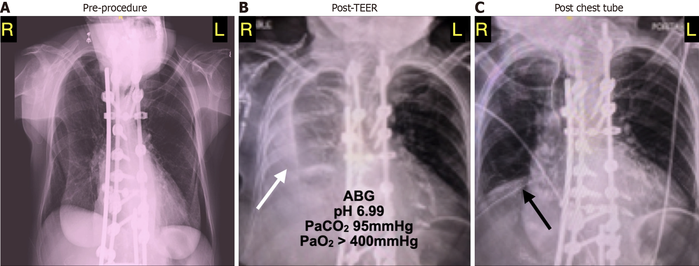
Figure 2 Chest X-ray.
A: Pre-procedure chest radiograph obtained the day before the scheduled mitral clip procedure. The lung fields appear hyperinflated with flattened diaphragms. There is extensive hardware (rods) seen along the spine; B: Post-procedure chest radiograph showing new haziness (white arrow) consistent with fluid. At this time, the patient was hypotensive and severely acidotic most due to a respiratory acidosis presumably related to extensive right lung collapse; C: Post-chest-tube-placement chest radiograph showing clearance of right pleural fluid. The lung fields appear smaller perhaps consistent with pulmonary atelectasis. As the prior films also see, there is extensive hardware along the spine.
- Citation: Seidler N, Asher SR, Chen T, Gordon P, Sodha N, Maslow A. Low-pressure tamponade due to hemothorax after transcatheter edge-to-edge repair of the mitral valve. World J Cardiol 2025; 17(5): 106567
- URL: https://www.wjgnet.com/1949-8462/full/v17/i5/106567.htm
- DOI: https://dx.doi.org/10.4330/wjc.v17.i5.106567


