
















©The Author(s) 2025.
World J Cardiol. Dec 26, 2025; 17(12): 112126
Published online Dec 26, 2025. doi: 10.4330/wjc.v17.i12.112126
Published online Dec 26, 2025. doi: 10.4330/wjc.v17.i12.112126
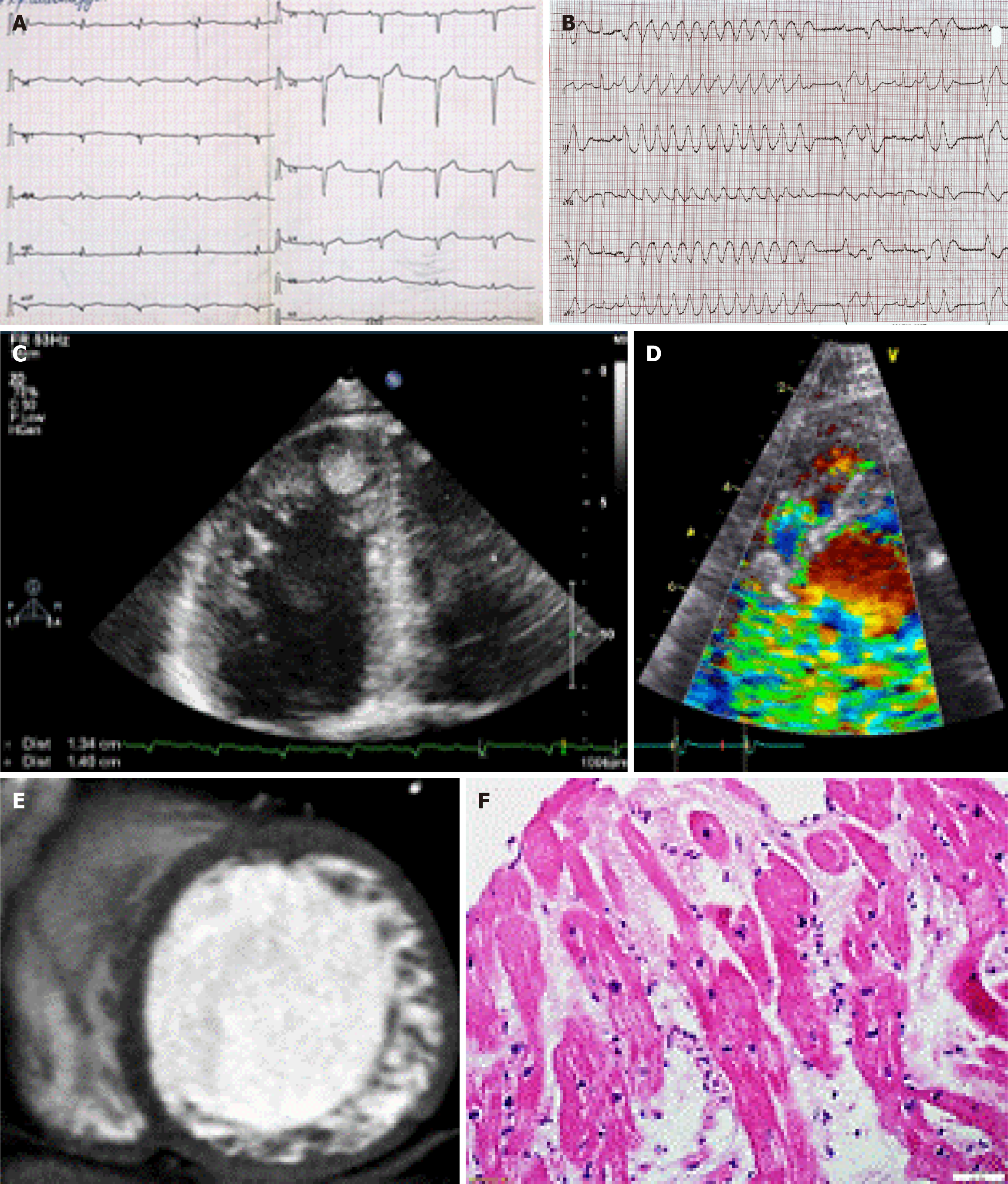
Figure 1 Visual, electrocardiographic, and morphological features of left ventricular noncompaction.
A and B: Electrocardiogram indicated low QRS voltage and non-sustained ventricular tachycardia; C and D: Echocardiogram indicated left ventricular noncompaction including a thrombus in the apex of left ventricle and intertrabecular blood flow by Doppler; E: Cardiac computed tomography indicated that the ratio of noncompact and compact myocardium was more than 2.3; F: Endomyocardial biopsy of the right ventricle. Hematoxylin and eosin staining indicated lymphocytic infiltration and necrosis as a sign of myocarditis.
- Citation: Blagova OV, Varionchik NV, Pavlenko EV, Sedov VP, Lutokhina YA. Predictors of life-threatening events in adult patients with left ventricular noncompaction. World J Cardiol 2025; 17(12): 112126
- URL: https://www.wjgnet.com/1949-8462/full/v17/i12/112126.htm
- DOI: https://dx.doi.org/10.4330/wjc.v17.i12.112126