
















©The Author(s) 2025.
World J Cardiol. Dec 26, 2025; 17(12): 112047
Published online Dec 26, 2025. doi: 10.4330/wjc.v17.i12.112047
Published online Dec 26, 2025. doi: 10.4330/wjc.v17.i12.112047
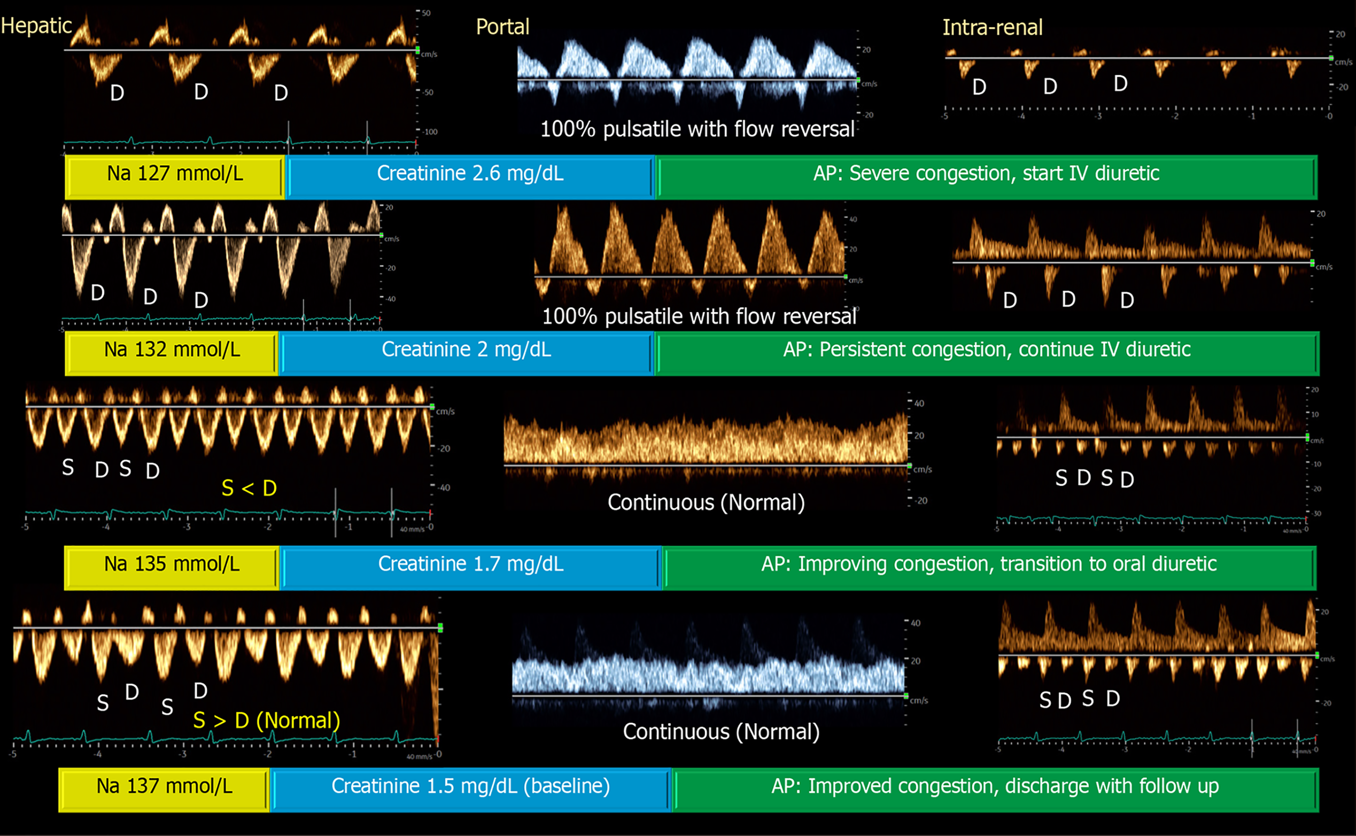
Figure 2 Venous excess ultrasound waveforms illustrate a case of acute kidney injury and hyponatremia, where serial assessment of venous Doppler patterns during daily clinical exams demonstrated progressive improvement with diuretic therapy[22].
Notably, the patient had no pedal edema or visible jugular venous distention and was initially presumed to be hypovolemic. However, point-of-care ultrasound revealed elevated right atrial pressure and severe venous congestion. The patient had heart failure with reduced ejection fraction and a chronically dilated inferior vena cava (IVC), making reliance on IVC diameter alone inadequate for follow-up evaluation. Citation: Koratala A, Ronco C, Kazory A. Multi-Organ Point-Of-Care Ultrasound in Acute Kidney Injury. Blood Purif 2022; 51: 967-971. Copyright© S. Karger AG, Basel 2022. Published by S. Karger AG, Basel. The authors have obtained the permission for figure using from the Springer Nature Publishing Group (Supplementary material).
- Citation: Chandra NC, Bhattacharya D, Koratala A. Point-of-care ultrasound: Uniting cardiology and nephrology at the bedside. World J Cardiol 2025; 17(12): 112047
- URL: https://www.wjgnet.com/1949-8462/full/v17/i12/112047.htm
- DOI: https://dx.doi.org/10.4330/wjc.v17.i12.112047