
















©The Author(s) 2025.
World J Cardiol. Dec 26, 2025; 17(12): 112047
Published online Dec 26, 2025. doi: 10.4330/wjc.v17.i12.112047
Published online Dec 26, 2025. doi: 10.4330/wjc.v17.i12.112047
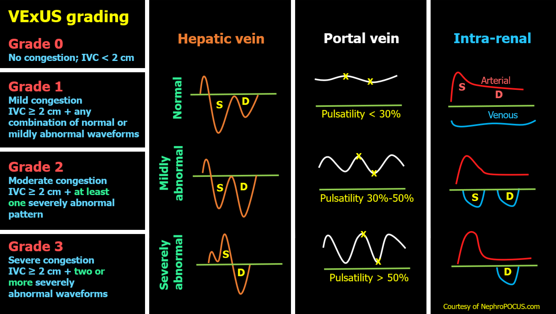
Figure 1 Venous excess ultrasound Grading System: When the inferior vena cava diameter exceeds 2 cm, further evaluation of the hepatic, portal, and intrarenal venous Doppler waveforms is recommended.
Abnormalities in these waveforms correlate with the degree of venous congestion. Hepatic vein Doppler is classified as mildly abnormal when the S wave is smaller than the D wave but remains below the baseline, and as severely abnormal when the S wave is reversed. Portal vein Doppler is mildly abnormal with 30%-50% pulsatility and severely abnormal when pulsatility is 50% or greater. Intrarenal vein Doppler is mildly abnormal when pulsatile with distinct S and D components, and severely abnormal when monophasic with a continuous D-only pattern. This figure was adapted from NephroPOCUS.com with permission. The corresponding author, Dr. Abhilash Koratala, is the website owner and copyright holder. Available from: https://nephropocus.com/2021/10/05/vexus-flash-cards/.
- Citation: Chandra NC, Bhattacharya D, Koratala A. Point-of-care ultrasound: Uniting cardiology and nephrology at the bedside. World J Cardiol 2025; 17(12): 112047
- URL: https://www.wjgnet.com/1949-8462/full/v17/i12/112047.htm
- DOI: https://dx.doi.org/10.4330/wjc.v17.i12.112047