Copyright
©The Author(s) 2022.
World J Cardiol. Sep 26, 2022; 14(9): 508-513
Published online Sep 26, 2022. doi: 10.4330/wjc.v14.i9.508
Published online Sep 26, 2022. doi: 10.4330/wjc.v14.i9.508
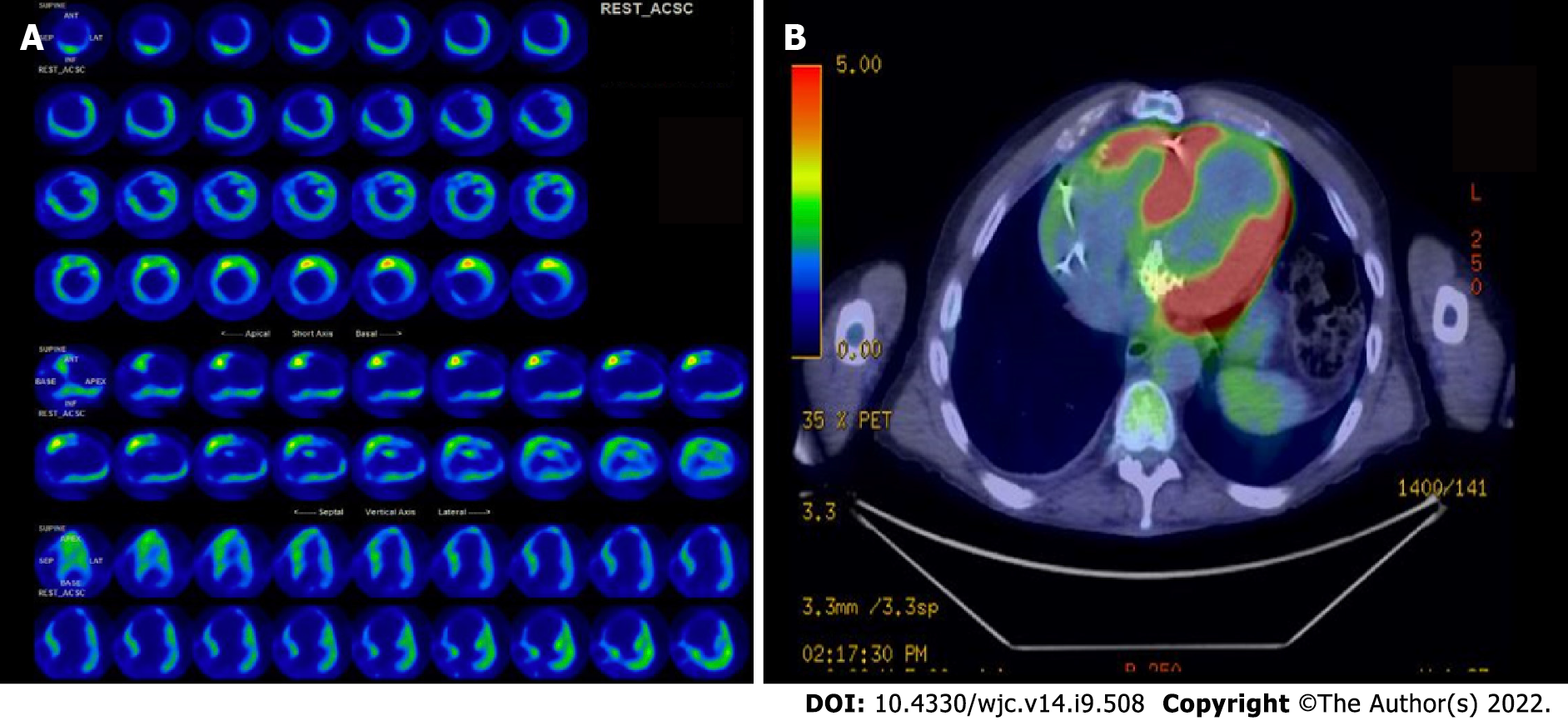
Figure 1 18-Flourine fluorodeoxyglucose positron emission tomography scan.
A: Heterogenous areas of increased uptake involving septal, lateral as well as basal and anterior wall of left ventricle suggestive of myocarditis; B: Whole body positron emission tomography obtained after 1 mo with focus on cardiac structure showing no evidence of residual myocarditis.
- Citation: Goyal A, Dalia T, Bhyan P, Farhoud H, Shah Z, Vidic A. Rare case of chronic Q fever myocarditis in end stage heart failure patient: A case report. World J Cardiol 2022; 14(9): 508-513
- URL: https://www.wjgnet.com/1949-8462/full/v14/i9/508.htm
- DOI: https://dx.doi.org/10.4330/wjc.v14.i9.508


