Copyright
©The Author(s) 2022.
World J Cardiol. Jun 26, 2022; 14(6): 372-381
Published online Jun 26, 2022. doi: 10.4330/wjc.v14.i6.372
Published online Jun 26, 2022. doi: 10.4330/wjc.v14.i6.372
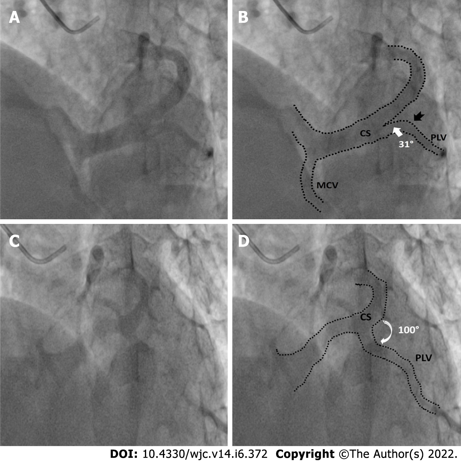
Figure 3 Favourable and unfavourable take off angles of posterolateral vein.
A: Example of posterolateral vein (PLV) with favourable angle-fluoroscopic image; B: Corresponding rendered image with dotted lines outlining coronary sinus (CS) morphology, white arrow denoting narrow angle between body of CS and PLV, black notched arrow represents single bend in PLV; C: Example of unfavourable angle PLV-fluoroscopic image; D: Corresponding rendered image with dotted lines outlining CS morphology, white curved arrow denoting wide angle between PLV and CS body.
- Citation: Pradhan A, Bajaj V, Vishwakarma P, Bhandari M, Sharma A, Chaudhary G, Chandra S, Sethi R, Narain VS, Dwivedi S. Study of coronary sinus anatomy during levophase of coronary angiography. World J Cardiol 2022; 14(6): 372-381
- URL: https://www.wjgnet.com/1949-8462/full/v14/i6/372.htm
- DOI: https://dx.doi.org/10.4330/wjc.v14.i6.372


