Copyright
©The Author(s) 2021.
World J Cardiol. Oct 26, 2021; 13(10): 593-598
Published online Oct 26, 2021. doi: 10.4330/wjc.v13.i10.593
Published online Oct 26, 2021. doi: 10.4330/wjc.v13.i10.593
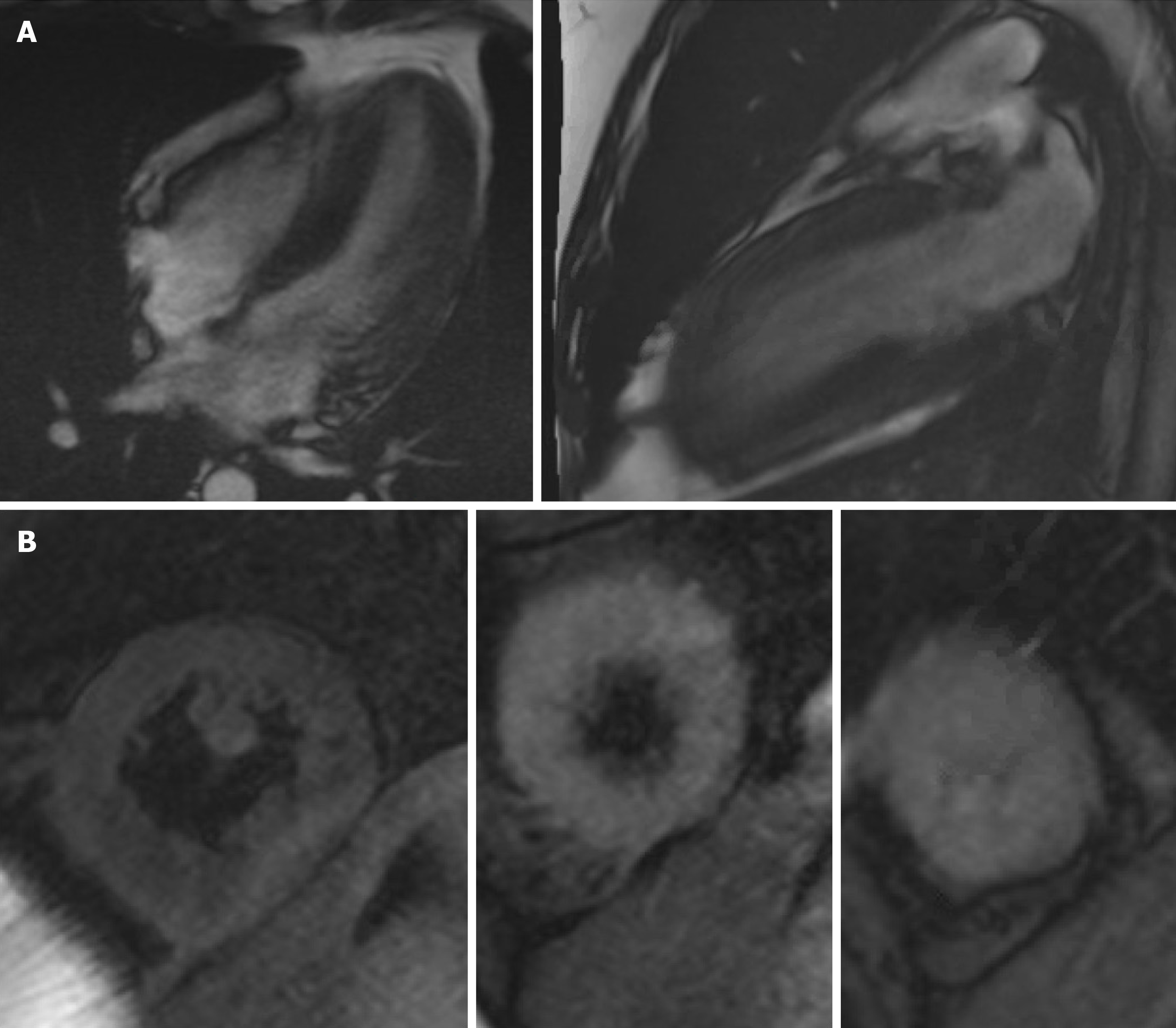
Figure 3 Cardiac magnetic resonance imaging.
A: 4 chamber cine, 2 chamber cine showing EF of 40% with global hypokinesis; B: T2 Weighted images showing edema in mid and apical segments.
- Citation: Jolly G, Dacosta Davis S, Ali S, Bitterman L, Saunders A, Kazbour H, Parwani P. Cardiac involvement in hydrocarbon inhalant toxicity — role of cardiac magnetic resonance imaging: A case report. World J Cardiol 2021; 13(10): 593-598
- URL: https://www.wjgnet.com/1949-8462/full/v13/i10/593.htm
- DOI: https://dx.doi.org/10.4330/wjc.v13.i10.593


