Copyright
©The Author(s) 2020.
World J Cardiol. Sep 26, 2020; 12(9): 437-449
Published online Sep 26, 2020. doi: 10.4330/wjc.v12.i9.437
Published online Sep 26, 2020. doi: 10.4330/wjc.v12.i9.437
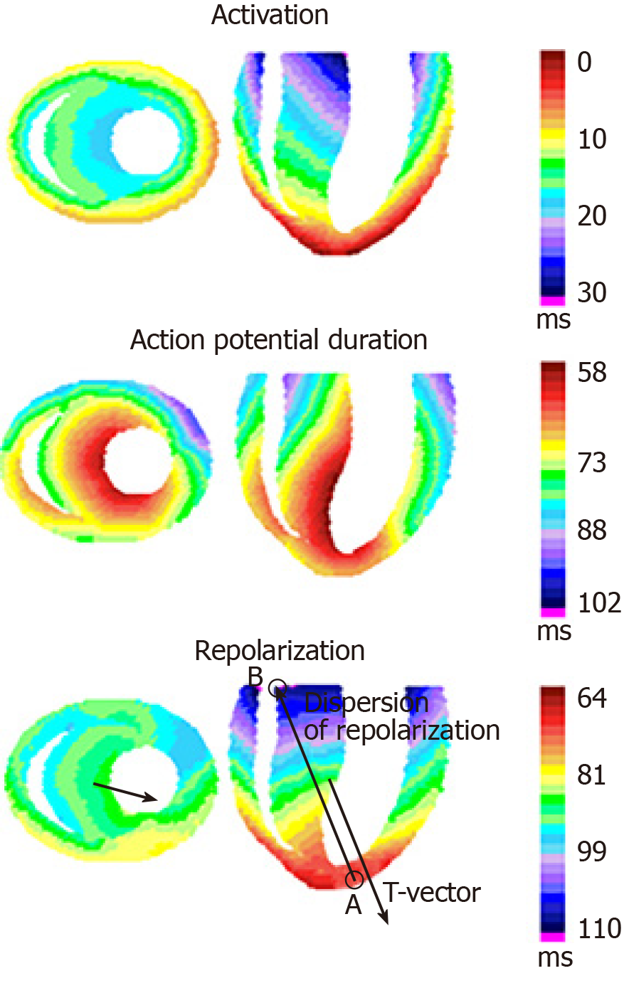
Figure 2 Realistic activation sequence, action potential duration distribution and end-of-repolarization sequence in the rabbit heart ventricles’ model, simulated from intramural and epicardial measurements[56].
The middle transversal and longitudinal cross-sections of the model and the corresponding T-vector projections are shown. Transmural, apicobasal, anterior-posterior and left-to-right gradients in activation times, action potential duration and end-of-repolarization times, reconstructed from experimental data, are presented. A and B – the regions of the earliest and the latest end-of-repolarization, correspondingly. T-vector direction is parallel but opposite to the general direction of repolarization sequence.
- Citation: Arteyeva NV. Dispersion of ventricular repolarization: Temporal and spatial. World J Cardiol 2020; 12(9): 437-449
- URL: https://www.wjgnet.com/1949-8462/full/v12/i9/437.htm
- DOI: https://dx.doi.org/10.4330/wjc.v12.i9.437


