Copyright
©The Author(s) 2018.
World J Cardiol. Sep 26, 2018; 10(9): 97-109
Published online Sep 26, 2018. doi: 10.4330/wjc.v10.i9.97
Published online Sep 26, 2018. doi: 10.4330/wjc.v10.i9.97
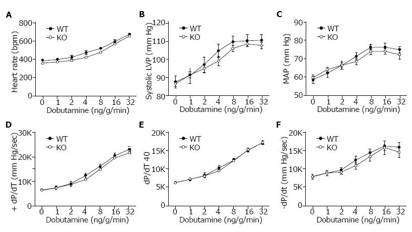
Figure 3 Cardiovascular performance in WT and KO mice.
Intraventricular and intra-arterial pressure measurements were recorded using transducers in the left ventricle and right femoral artery of anesthetized 2-3 mo old WT (Slc4a4flx/flx) and KO (Slc4a4flx/flx(Cre)) mice under both basal conditions and in response to β-adrenergic stimulation (intravenous infusion of increasing doses of dobutamine). A: Heart rate; B: Systolic left ventricular pressure; C: Mean arterial pressure; D: Positive dP/dt in mm Hg/sec; E: Positive dP/dt at 40 mm Hg; F: Negative dP/dt in mm Hg/sec is shown for WT and KO mice. n = 8 WT (4 female, 4 male) and 8 KO (4 female, 4 male) mice.
- Citation: Vairamani K, Prasad V, Wang Y, Huang W, Chen Y, Medvedovic M, Lorenz JN, Shull GE. NBCe1 Na+-HCO3- cotransporter ablation causes reduced apoptosis following cardiac ischemia-reperfusion injury in vivo. World J Cardiol 2018; 10(9): 97-109
- URL: https://www.wjgnet.com/1949-8462/full/v10/i9/97.htm
- DOI: https://dx.doi.org/10.4330/wjc.v10.i9.97


