Copyright
©The Author(s) 2017.
World J Cardiol. May 26, 2017; 9(5): 466-469
Published online May 26, 2017. doi: 10.4330/wjc.v9.i5.466
Published online May 26, 2017. doi: 10.4330/wjc.v9.i5.466
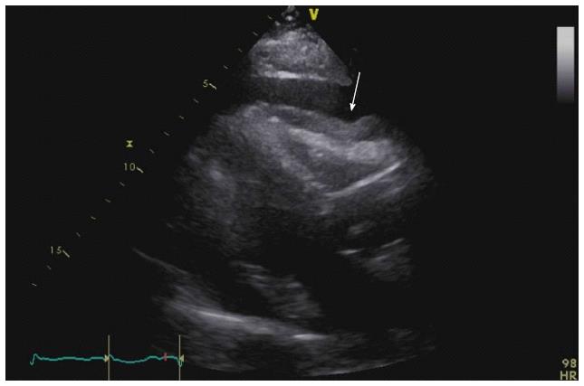
Figure 1 Transthoracic echocardiogram showing a large pericardial effusion with right ventricular diastolic indentation and collapse suggestive of tamponade.
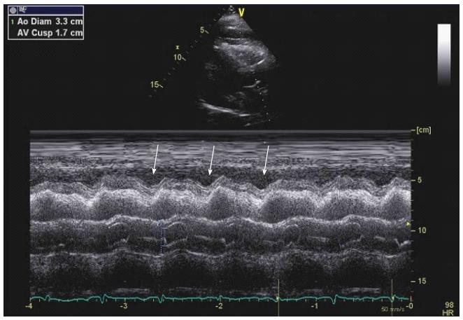
Figure 2 M-mode showing right ventricular diastolic indentation.
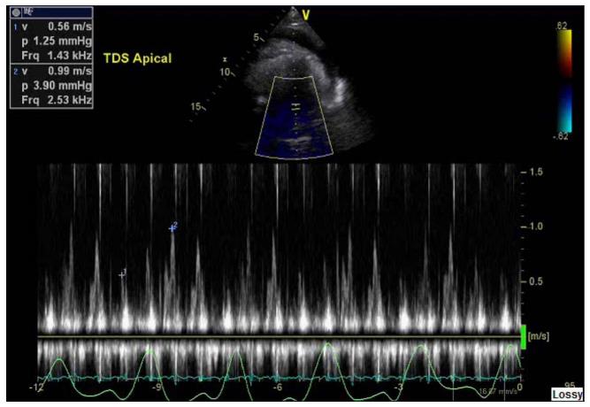
Figure 3 Transthoracic echocardiogram showing a mitral valve inflow E wave velocity greater than 25% respiratory variation suggestive of tamponade physiology.
- Citation: Ramirez R, Lasam G. Cough induced syncope: A hint to cardiac tamponade diagnosis. World J Cardiol 2017; 9(5): 466-469
- URL: https://www.wjgnet.com/1949-8462/full/v9/i5/466.htm
- DOI: https://dx.doi.org/10.4330/wjc.v9.i5.466


