Copyright
©2014 Baishideng Publishing Group Co.
World J Cardiol. Apr 26, 2014; 6(4): 154-174
Published online Apr 26, 2014. doi: 10.4330/wjc.v6.i4.154
Published online Apr 26, 2014. doi: 10.4330/wjc.v6.i4.154
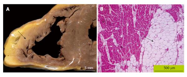
Figure 1 Typical pathology findings in arrhythmogenic ventricular cardiomyopathy/dysplasia.
A: Macroscopic finding in a patient with arrhythmogenic right ventricular cardiomyopathy/dysplasia (ARVC/D). The myocardium of the right ventricular free wall is partially replaced by fibro-fatty tissue (black arrow) that typically begins in the epicardial region and at later stages expands transmurally; B: Endomyocardial biopsy from a patient with ARVC/D demonstrating fatty (black arrow) replacement of the right ventricular myocardium. Strands of myocardium are still visible (white arrow, heidenhain trichrome, magnification × 60).
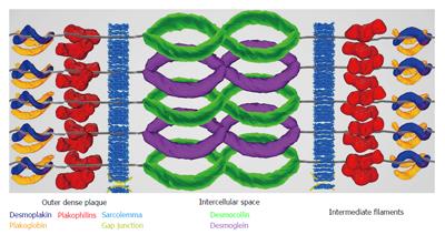
Figure 2 Molecular model of the desmosome: in the desmosomal complex the intermediate filaments of the cytoskeleton (desmin in the heart) are linked to the transmembranous cadherins (desmocollin and desmoglein) via armadillo proteins (plakoglobin and plakophilin) and desmoplakin.
This interaction is crucial for myocardial mechanical and electrical stability. Mutations in arrhythmogenic right ventricular cardiomyopathy mostly affect desmosomal proteins.
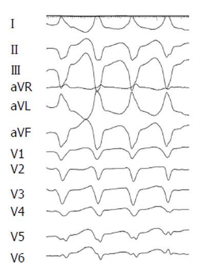
Figure 3 Monomorphic sustained ventricular tachycardia with left bundle branch block morphology and superior axis (II, III, aVF negative), a major criterion for arrhythmogenic right ventricular cardiomyopathy/dysplasia according to the revised 2010 task force criteria.
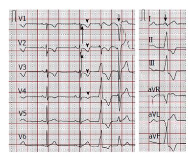
Figure 4 Electrocardiographic findings.
A 12-lead surface electrocardiogram (25 mm/s, 10 mm/mV) showing typical depolarization abnormalities (prolonged terminal activation duration in V1-V2, a minor criterion according to 2010 task force criteria, long arrows) and repolarization abnormalities (T-wave inversions V1-V4 in the absence of complete right bundle branch block, a major criterion according to 2010 task force criteria, arrowheads), and premature ventricular contractions with two different morphologies (short arrows).
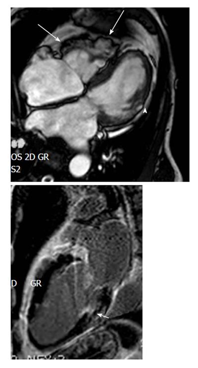
Figure 5 Regional right ventricular dyskinesia of the right free wall detected by cardiac imaging are considered as a major criterion for right ventricular cardiomyopathy/dysplasia according to the revised task force criteria if additionally right ventricle dilation or impaired right ventricle ejection fraction are present.
These cardiac magnetic resonance images (upper panel 4-chamber view, lower panel 2-chamber view late sequences) show aneurysms of the RV free wall (long arrows), and LV involvement detected by a small akinetic region (arrowhead) and late gadolinium enhancement of the posterior LV wall (short arrow), confirming biventricular involvement. ARVC/D: arrhythmogenic right ventricular cardiomyopathy/dysplasia; RV: Right ventricle; LV: Left ventricle.
- Citation: Saguner AM, Brunckhorst C, Duru F. Arrhythmogenic ventricular cardiomyopathy: A paradigm shift from right to biventricular disease. World J Cardiol 2014; 6(4): 154-174
- URL: https://www.wjgnet.com/1949-8462/full/v6/i4/154.htm
- DOI: https://dx.doi.org/10.4330/wjc.v6.i4.154


