Copyright
©The Author(s) 2021.
World J Cardiol. Dec 26, 2021; 13(12): 654-675
Published online Dec 26, 2021. doi: 10.4330/wjc.v13.i12.654
Published online Dec 26, 2021. doi: 10.4330/wjc.v13.i12.654
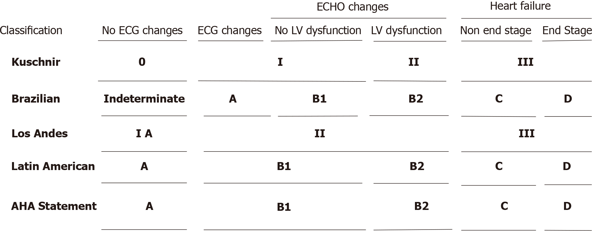
Figure 1 Schematic representation of the different classification systems of Chagas disease.
We assumed that patients with an enlarged heart on chest radiography would have left ventricular systolic dysfunction on echocardiography in order to be able to compare all classifications.
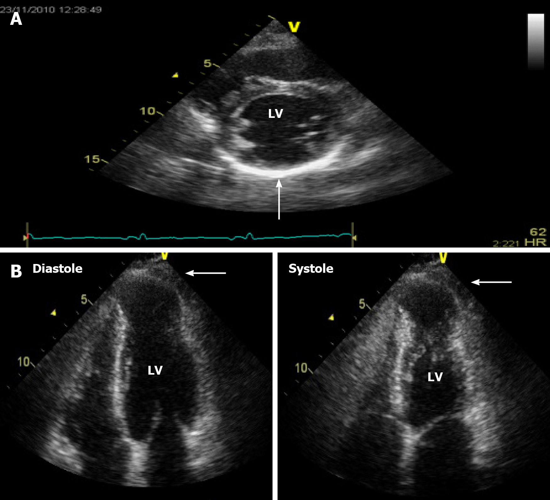
Figure 2 Note the hyperrefringent akinetic area in the basal inferolateral left ventricular wall (A) and the left ventricular apical aneurysm in the diastolic and systolic left ventricular images (B).
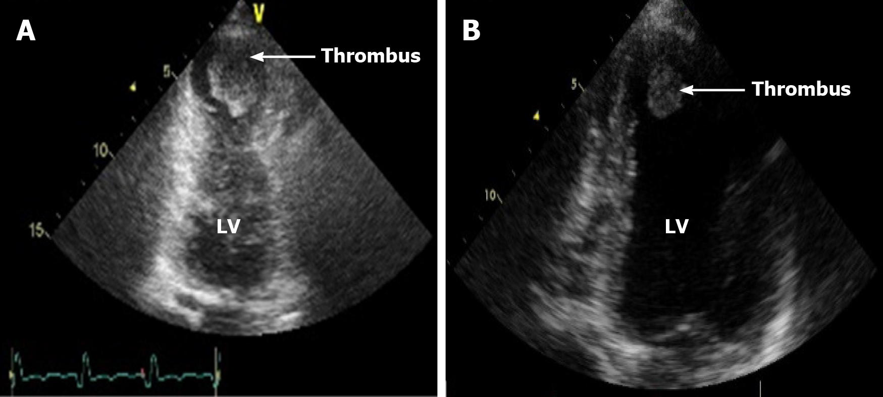
Figure 3 Note the thrombi in the apical location in a patient with a large apical aneurysm (A) and a patient with severe left ventricular dilation and dysfunction (B).
Figure 4 Cardiac magnetic resonance imaging of a patient with stage B1 Chagas heart disease.
A: Myocardial delayed enhancement on 2-chamber apical slice depicts areas of cardiac fibrosis in the apical segments and an apical thrombus; B: Myocardial delayed enhancement protocol on left ventricular short-axis slices depicts areas of cardiac fibrosis in the apical and basal left ventricular walls of a patient at stage B1 of Chagas heart disease.
- Citation: Saraiva RM, Mediano MFF, Mendes FS, Sperandio da Silva GM, Veloso HH, Sangenis LHC, Silva PSD, Mazzoli-Rocha F, Sousa AS, Holanda MT, Hasslocher-Moreno AM. Chagas heart disease: An overview of diagnosis, manifestations, treatment, and care. World J Cardiol 2021; 13(12): 654-675
- URL: https://www.wjgnet.com/1949-8462/full/v13/i12/654.htm
- DOI: https://dx.doi.org/10.4330/wjc.v13.i12.654


