INTRODUCTION AND EDUCATIONAL EXPERIENCE
Since I was young, I (Figure 1) have been interested in being a teacher, and that feeling grew and consolidated along with my professional journey. The scientific world was introduced to me during high school. From 1990 to 1994, I studied the Biotechnology course in a reputable Federal Institution from Rio de Janeiro State, Brazil, called Escola Técnica Federal de Química - ETFQ (currently CEFETEQ), an excellent technical school. Over those years, the disciplines related to the Microbiology area (Bacteriology, Mycology, Virology, Protozoology and Immunology) and the laboratory classes produced a great curiosity, motivation and stimulation of scientific thought, which ignited my desire to be a scientist. With this proposal in mind, in 1994, I started my bachelor degree in the Microbiology and Immunology course at the Federal University of Rio de Janeiro (UFRJ), being one of the 35 students approved to constitute the first class of that novel graduation course. In parallel, I worked as a Biotechnology technician at the Biochemistry Department of the State University of Rio de Janeiro (UERJ) under the supervision of Dr. Claudia Vitória de Moura Gallo, an exemplar professional and an excellent person, who contributed notably to turn my dream into reality. In early 1999, I finished the undergraduate program and started a Master’s degree at the Institute of Microbiology Prof. Paulo de Góes (IMPPG)-UFRJ. During the period from mid 2000 until early 2002, I developed my doctoral thesis at the IMPPG-UFRJ under the supervision of Dr. Rosangela Maria de Araújo Soares. Since August 2002, I have been Professor at the Department of General Microbiology of the IMPPG-UFRJ and, since then, I have been teaching lessons to several undergraduate courses including Microbiology and Immunology, Nursing, Biology and Pharmacy. Still, I effectively participate in two postgraduate courses at UFRJ: Microbiology from IMPPG and Biochemistry from Chemistry Institute.
Figure 1 André Luis Souza dos Santos, Associate Professor, Laboratory of Multidisciplinary Studies on Microbial Biochemistry, Department of General Microbiology, Institute of Microbiology Prof.
Paulo de Góes, Federal University of Rio de Janeiro, Rio de Janeiro, RJ 21941-902, Brazil.
Indubitably, my professional work has only been fully developed because I have a research group consisting of competent professionals, including technicians and undergraduate, masters, doctoral and postdoctoral students, who are extremely dedicated and committed to scientific thinking. I would like take this opportunity to express and reiterate my full admiration and gratitude to all my students. I would also like to thank to the several Brazilian researchers who have contributed immensely to my work, in particular Dr. Marta Helena Branquinha (IMPPG-UFRJ), Dr. Eliana Barreto-Bergter (IMPPG-UFRJ), Dr. Lucy Seldin (IMPPG-UFRJ), Dr. Celuta Sales Alviano (IMPPG-UFRJ), Dr. Claudia Masini d’Avila-Levy (Fundação Oswaldo Cruz-FIOCRUZ) and Dr. Lucimar Ferreira Kneipp (FIOCRUZ). My research has been supported by the Brazilian agencies: Conselho Nacional de Desenvolvimento Científico e Tecnológico (CNPq), Conselho de Ensino para Graduados e Pesquisas (CEPG/UFRJ), Fundação de Amparo à Pesquisa do Estado do Rio de Janeiro (FAPERJ), Fundação Universitária José Bonifácio (FUJB) and Coordenação de Aperfeiçoamento de Pessoal de Nível Superior (CAPES). I have also been supported by a CNPq fellowship since 2005 and by a FAPERJ fellowship since 2007.
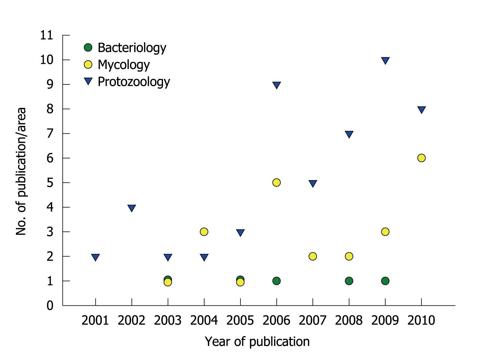
Over the past 10 years: (1) I supervised 16 monographs of graduate students, 10 master theses and 4 doctoral theses; (2) I published 79 papers in the field of Bacteriology (n = 5), Mycology (n = 22) and Protozoology (n = 52) (Figure 2); and (3) I was invited to participate as a speaker at national and international meetings. I am a peer reviewer for international scientific journals, as well as career and research grant committees. In addition, I have accepted invitations to write reviews and book chapters on the themes: (1) relevance of proteolytic enzymes produced by microorganisms; and (2) antimicrobial properties of protease inhibitors[1-11].
Figure 2 Publication of scientific papers by the research group led by André Santos.
The graphic summarizes the numbers and specific areas of Microbiology in relation to papers published during the past ten years.
ACADEMIC STRATEGIES AND GOALS
Our work group is distinguished by its multidisciplinary nature, with direct involvement of different research institutions from Brazil (other Departments and Institutes from UFRJ, UERJ, FIOCRUZ, Universidade Federal Fluminense (UFF), Universidade Federal do Estado do Rio de Janeiro (UNIRIO), Universidade do Estado de São Paulo (USP), Universidade Federal de São Paulo (UNIFESP), Universidade Federal do Espírito Santo (UFES)) and from other countries, generating productive and effective collaborations. Several publications in high-ranked journals, e.g. FEMS Microbiology Reviews, PLoS One, Archives of Biochemistry and Biophysics, Journal of Antimicrobial Chemotherapy, Journal of Clinical Microbiology, International Journal of Antimicrobial Agents, Microbes and Infection, International Journal for Parasitology, Protist, Parasitology and Medical Mycology, were produced in collaboration with these partners.
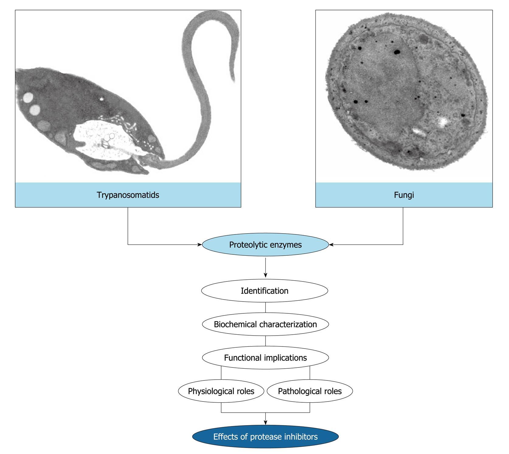
Over the last years, my group has focused on the identification, biochemical characterization and discovery of biological functions of proteases produced by microorganisms, with emphasis in trypanosomatids and fungi (Figure 3). More recently, we have started to study protease inhibitors in an attempt to use these bioactive compounds as a new therapeutic proposal against eukaryotic pathogenic microorganisms (Figure 3).
Figure 3 Rationale of the research works developed in the André Santos’ laboratory.
The main purpose of our study focuses on the identification and biochemical characterization of cellular and/or extracellular proteases produced by eukaryotic microorganisms, especially trypanosomatids and fungi. Subsequently, we have focused on the discovery of possible biological functions for these hydrolytic enzymes in both the social context of the microbial cell and the participation in interaction events with biotic and abiotic substrates. Finally, we have used the protease inhibitors in an attempt to block vital processes in microbial cells, thus preventing a successful infection.
RESEARCH ACHIEVEMENTS
Proteolytic enzymes and their inhibitors: an overview
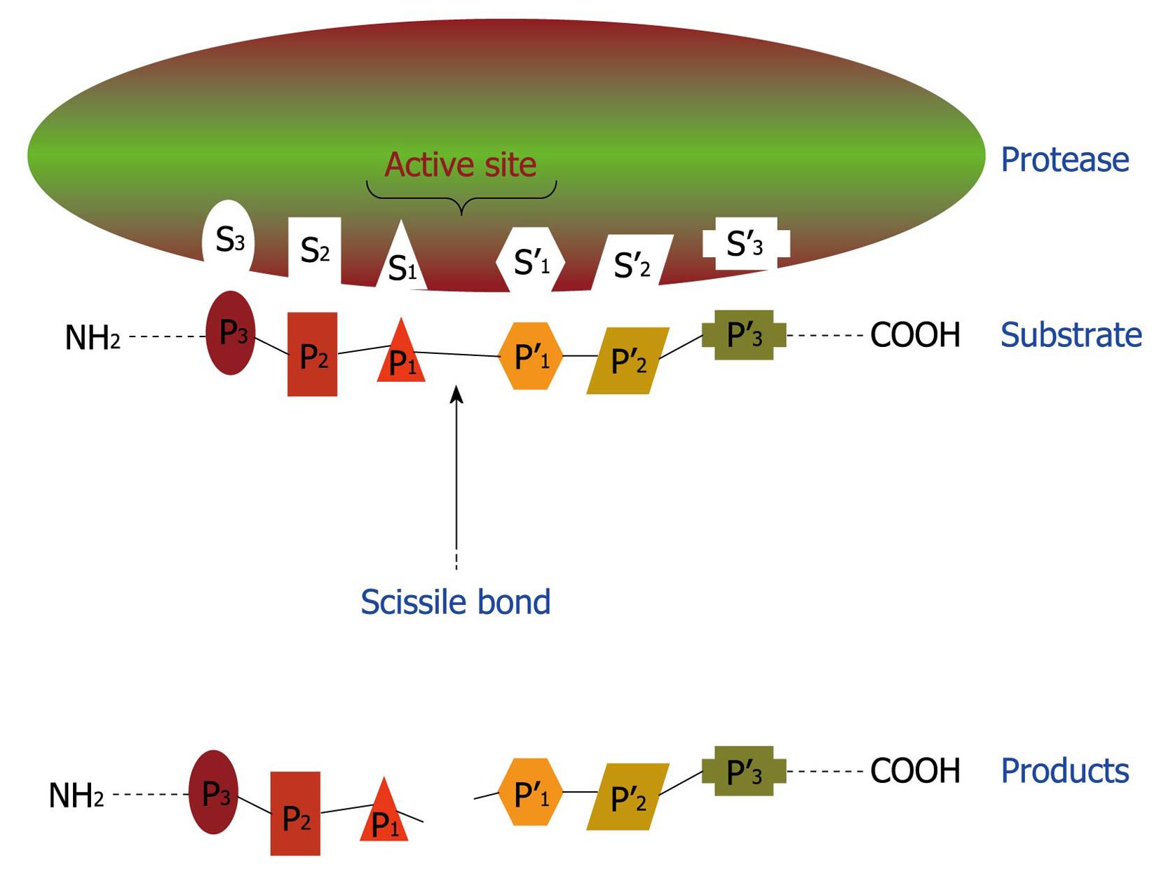
Proteolytic enzymes catalyze the cleavage of peptide bonds, which link amino acid residues in proteins and peptides. A redundant set of terms is used by the scientific community to refer to proteolytic enzymes, including: peptide hydrolase, peptidase and protease. All proteases bind their substrates in a groove or cleft, where peptide bond hydrolysis occurs (Figure 4). Amino acid side chains of substrates occupy proteolytic enzyme sub-sites in the groove, designated as S3, S2, S1, S1’, S2’, S3’, that bind to corresponding substrate/inhibitor residues P3, P2, P1, P1’, P2’, P3’ with respect to the cleavable peptide bond (Figure 4). After the proteinaceous substrate cleavage, at least two smaller peptides can be generated (Figure 4)[12-15].
Figure 4 Schematic representation of binding region and catalytic site of a protease.
This hypothetical protease possesses six subsites (S1-S3 and S1’-S3’) in its catalytic site and, consequently, is able to recognize and bind to a sequence of six amino acids (P1-P3 and P1’-P3’) in the proteinaceous substrate. After proteolysis, at least two smaller peptides are generated as the reaction products.
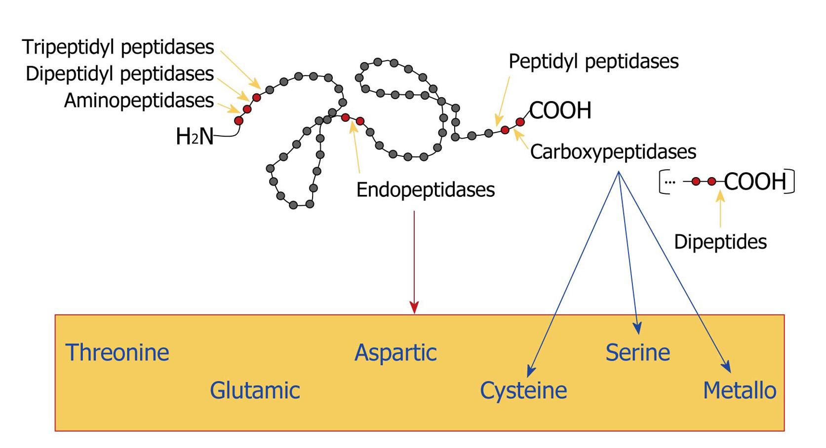
Proteases are subdivided into two major groups depending on their site of action: exopeptidases and endopeptidases. Exopeptidases cleave the peptide bond proximal to the amino (NH2) or carboxy (COOH) termini of the proteinaceous substrate, whereas endopeptidases cleave peptide bonds within a polypeptide chain. Based on their site of action at the NH2 terminal, the exopeptidases are classified as aminopeptidases, dipeptidyl peptidases or tripeptidyl peptidases that act at a free NH2 terminus of the polypeptide chain and liberate a single amino acid residue, a dipeptide or a tripeptide, respectively. Carboxypeptidases or peptidyl peptidases act at the COOH terminal of the polypeptide chain and liberate a single amino acid or a dipeptide (which can be hydrolyzed by the action of a dipeptidase). Carboxypeptidases can be further divided into three major groups: serine, metallo and cysteine carboxypeptidases, based on the functional group present at the active site of the enzymes. Similarly, endopeptidases are classified according to essential catalytic residues at their active sites in: serine, metallo, glutamic, threonine, cysteine and aspartic endopeptidases (Figure 5). Conversely, there are a few miscellaneous proteases that do not precisely fit into the standard classification[12-15].
Figure 5 Classification of proteases.
Gray circles represent amino acids and red circles indicate the amino acid sequence that is bound to the proteolytic enzyme. Yellow arrows point to the site of cleavage. The blue arrows indicate the classification of carboxypeptidases and the red arrow shows the box containing all the classes of endopeptidases, according to the chemical group present in their catalytic sites.
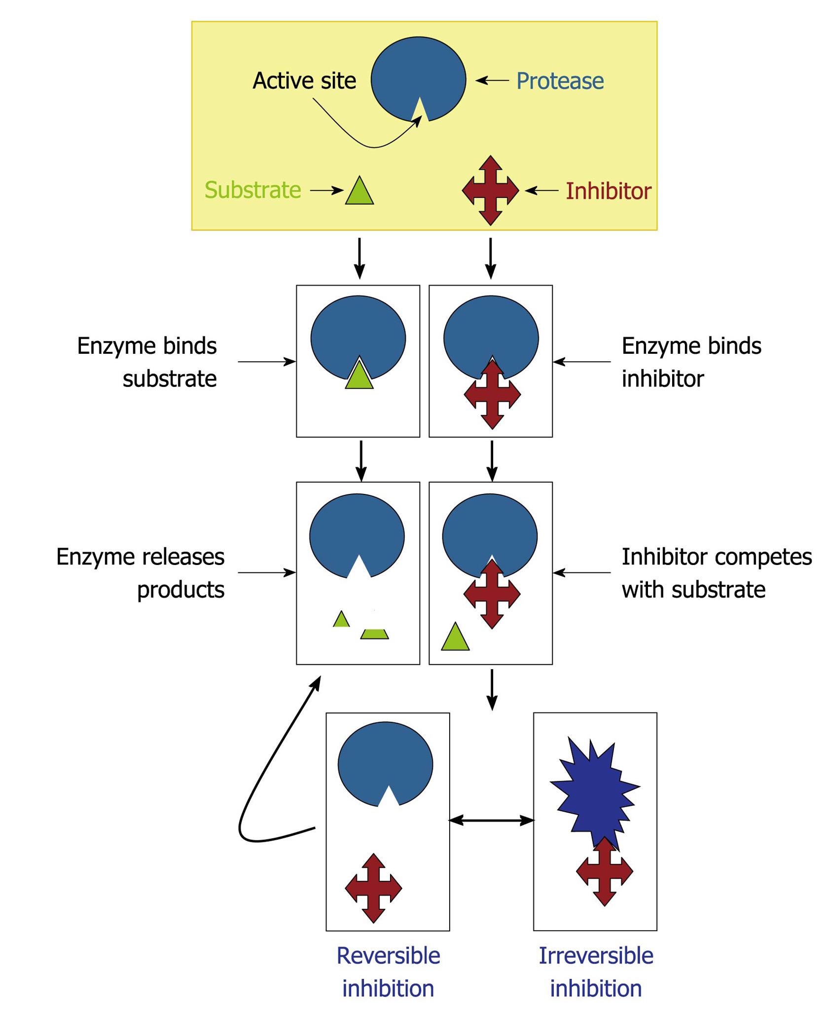
The class of a protease is characteristically determined according to the effects of proteolytic inhibitors on the enzymatic activity[16,17]. Protease inhibitors enter or block a protease active site to prevent substrate access. In competitive inhibition, the inhibitor binds to the active site, thus preventing enzyme-substrate interaction. In non-competitive inhibition, the inhibitor binds to an allosteric site, which alters the active site and makes it inaccessible to the substrate[16,17]. The proteolytic inhibitors can be divided into two functional classes on the basis of their interaction with the target protease: (1) irreversible trapping reactions and (2) reversible tight-binding reactions (Figure 6). Inhibitors which bind through a trapping mechanism change conformation after cleaving an internal peptide bond and “trap” the enzyme molecule covalently; neither the inhibitor nor protease can participate in further reactions. In tight-binding reactions, the inhibitor binds directly to the active site of the protease; these reactions are reversible and the inhibitor can dissociate from the proteolytic enzyme in either the virgin state, or after modification by the protease. Based on their structural dichotomy, proteolytic inhibitors can be generally classified into two large groups: low molecular mass peptidomimetic inhibitors and protein protease inhibitors composed of one or more peptide chains. Proteolytic inhibitors can be further classified into five groups (metallo, serine, threonine, cysteine and aspartic protease inhibitors) according to the mechanism employed at the active site of proteolytic enzymes they inhibit. Some proteolytic inhibitors interfere with more than one type of protease[16,17].
Figure 6 Mechanisms of protease inhibition.
The protease inhibitor competes with the substrate to bind to the active site of a protease and two distinct possibilities arise: (1) substrate binds to the catalytic site and then is cleaved by the protease, which releases the products or (2) inhibitor binds to the active site and by steric hindrance blocks the substrate attachment. In this last case, the inhibitor can promote an irreversible (the conformational structure of the protease is completely lost) or reversible inhibition (when the inhibitor disconnects from the enzyme, the substrate can bind to it).
Proteases produced by microorganisms: global functions
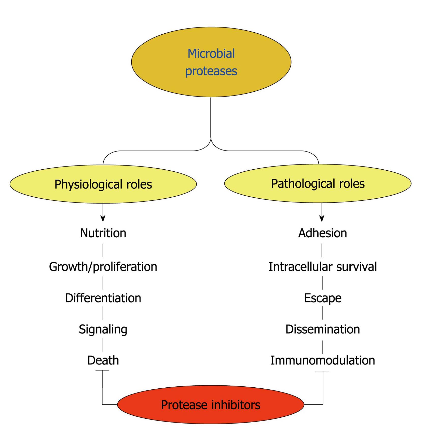
Proteases are essential for all life forms. They are involved in a multitude of physiological reactions from simple digestion of proteins for nutrition purposes to highly-regulated metabolic cascades (e.g. proliferation and growth, differentiation, signaling and death pathways), being essential factors for homeostatic control in both prokaryote and eukaryote cells (Figure 7)[12]. Proteases are also essential molecules in viruses, bacteria, fungi and protozoa for their colonization, invasion, dissemination and evasion of host immune responses, mediating and sustaining the infectious disease process (Figure 7). Collectively, proteases participate in different steps of the multifaceted interaction events between microorganism and host structures, being considered as virulent attributes. Consequently, the biochemical characterization of these proteolytic enzymes is of interest not only for understanding proteases in general but also for understanding their roles in microbial infections and thus their exploitation as targets for rational chemotherapy of microbial diseases[3,6,10,18-24].
Figure 7 Possible functions played by microbial proteases.
Surface and/or secreted proteases are able to cleave different host components such as serum proteins, antimicrobial peptides, surface molecules and structural proteinaceous compounds. The degradation of host proteins can help the microorganisms in several steps of their life cycle and pathogenesis including dissemination, adhesion, escape, nutrition and immunomodulation of the host immune response. These proteases can also contribute to maintaining basic metabolic processes in a microbial cell, which govern crucial events like proliferation, differentiation, signaling and death pathways. Proteolytic inhibitors are able to block one or several of these fundamental events.
Antimicrobial properties of proteolytic inhibitors
Current therapy for both fungal and trypanosomatid infections is suboptimal due to toxicity of the available therapeutic agents and the emergence of drug resistance[25-28]. Compounding these problems is the fact that many endemic countries and regions are economically poor. For that reason, the development of novel antifungal and/or anti-trypanosomatidal drugs is an imperative requirement. A number of new strategies to obstruct fungal/trypanosomatid biological processes have emerged; one of them is focused on protease inhibition. Currently, the main approach has been to obtain good inhibitors of the target protease, in the belief that inhibition of the activity will be therapeutic. In this context, our research group has published some works that corroborate this premise[1-6,10,29-39].
Aspartic protease inhibitors used in anti-human immunodeficiency virus therapy present anti-microbial properties
Lessons from the yeast Candida albicans (C. albicans), the filamentous fungus Fonsecaea pedrosoi (F. pedrosoi) and the protozoan Leishmania amazonensis (L. amazonensis) are illustrated as follows.
C. albicans:C. albicans is both a successful commensal and pathogen of humans that can infect a broad range of body sites[40]. The transition from commensalism to parasitism requires a susceptible host, which includes individuals with humoral and/or cellular deficiencies as well as persons submitted to different immunosuppressive procedures. Candidiasis is the most common fungal infection diagnosed in humans[41-43]. Due to the emergence of pathogens resistant to conventional antifungals and the toxicity of some antimycotics, intense efforts have been made to develop more effective antifungal agents for clinical use[44-48]. The pathogenesis of C. albicans is multifactorial and different virulence attributes are important during the various stages of infection[20,21,49-55]. Secreted aspartic proteases (Saps) play a role in several infection stages of C. albicans, being the most important virulence factors expressed by this opportunistic fungus. Actually, C. albicans possesses ten different SAP genes (SAP1 to SAP10), which are expressed according to distinct environments and host conditions[56-60]. Therefore, Saps are potential targets for the development of novel anti-C. albicans drugs[1,2,34,35]. In this context, several groups have demonstrated that aspartic protease inhibitors, including pepstatin A and the first generation of protease inhibitors used in anti-human immunodeficiency virus (HIV) therapy (nelfinavir, saquinavir, ritonavir and indinavir), are able to restrain Sap activity (especially Sap1, Sap2 and Sap3) as well as arrest crucial events of C. albicans yeast cells such as proliferation and adhesion to both abiotic (e.g. plastic and acrylic substrates) and biotic structures (e.g. surface of different epithelial cell lineages)[61-72]. Our results showed that amprenavir[72] (unpublished data) and lopinavir (unpublished data), two HIV aspartic protease inhibitors of the second generation, significantly inhibited the hydrolytic activity of Sap2 and also blocked the yeasts into mycelia transformation, an essential step during the candidiasis pathogenesis. In addition, scanning electron microscopy revealed prominent ultrastructural alterations of yeast cells, which corroborated the inhibition of cellular division by these protease inhibitors. Several surface and/or secreted molecules have had their expression/production significantly diminished including (1) mannose- and sialic acid-rich surface glycoconjugates, which are directly involved in adhesive properties and biofilm formation; (2) sterol content, which controls the membrane fluidity; (3) secretion of lipases (e.g. esterases and phospholipases), which are related to the host membrane disruption; and (4) catalase activity, which reduces the ability of yeasts to escape from oxidative stress generated by hydrogen peroxide, for example, released by host phagocytes[72] (unpublished data). However, it is also important to note that the inhibitory effects of HIV protease inhibitors both in in vitro and in vivo experimental models were observed at concentrations (μmol/L range) much higher than those needed for HIV protease inhibition (nmol/L range). This probably reflects a much lower affinity of these drugs for Sap than that for HIV protease[31,34]. Another explanation is that, in contrast to the very small and structurally simplified HIV protease, Saps are larger and more complex[60,73]. They possess a relatively large active site which might be responsible for the broader substrate specificity and also their susceptibilities to distinct aspartic protease inhibitors[60]. Nevertheless, the above concentrations may be achieved under current highly active antiretroviral therapy (HAART) regimens both in the blood[31], in human saliva (at least for indinavir)[74] and in lungs (at least for lopinavir)[75]. In this sense, our group has showed that lopinavir at 10 mg/kg promoted a therapeutic effect in an experimental murine model of disseminated candidiasis, with an efficacy comparable to that of fluconazole, a recognized anti-candidal drug (unpublished results).
F. pedrosoi:Fonsecaea is a genus containing pigmented filamentous fungus isolated from soil, rotten wood and decomposing plant material. F. pedrosoi is one of the major causative agents of chromoblastomycosis, a post-traumatic and chronic infection of subcutaneous tissues in humid tropical areas specially South America and Japan[8,76-79]. F. pedrosoi is a valuable model in cell biology, since its life cycle comprises different morphological states that include reproduction structures (conidia) and fungal forms usually found in the saprophytic (mycelia) and parasitic stage (sclerotic bodies)[8]. The first report on protease production by F. pedrosoi was described by our group[80], which demonstrated that the pattern of protease production and secretion by F. pedrosoi conidial cells was closely dependent on the culture medium composition: metalloproteases were induced after cultivation in complex culture medium, while aspartic proteases were detected under chemically defined growth conditions. Mycelia[81] and sclerotic cells (unpublished results) of F. pedrosoi were also able to secrete aspartic-type proteases. The aspartic proteases produced by conidia and mycelia were capable of degrading relevant host serum proteins (e.g. IgG and albumin) as well as extracellular matrix components (e.g. laminin, fibronectin and collagen)[80,81]. For that reason, the extracellular hydrolytic enzymes produced by F. pedrosoi cells, such as proteases and lipases[82], could support the initial development of this fungus inside the host, and the existence of two biochemically distinct secreted proteases makes it possible to cover a wide range of host conditions. The effect of saquinavir, ritonavir, indinavir and nelfinavir on the secreted proteases of F. pedrosoi was evaluated[81,83]. These compounds inhibited the extracellular aspartic proteolytic activity produced by both conidial and mycelial forms in a dose-dependent manner. Nelfinavir was the best inhibitor of the aspartic protease activity secreted by conidia and mycelia, restraining the hydrolytic activities around 80% at 50 μmol/L. Interestingly, recent isolated strains of F. pedrosoi produced higher levels of extracellular protease activity when compared with a laboratory-adapted strain[81,83], suggesting that the production of secreted aspartic-type proteases may be stimulated by interaction with the host. HIV aspartic protease inhibitors and pepstatin A also arrested the growth of conidial forms as well as transformation into mycelia[83], an essential step during the F. pedrosoi life cycle and virulence[8]. Pepstatin A showed a significant inhibition of conidial viability even at low concentration (0.1 μmol/L); however, the HIV protease inhibitors were toxic only at high concentrations (ranging from 50 to 200 μmol/L). The synergistic action on proliferation behavior between nelfinavir (25 μmol/L) and amphotericin B (3 μg/mL), when both were used at sub-inhibitory concentrations, was also observed[83]. Interestingly, HIV protease inhibitors-treated conidial cells presented irreversible ultrastructural alterations, as shown by transmission electron microscopy images such as invaginations in the cytoplasmic membrane and withdrawal of the cytoplasmic membrane from within the cell wall, disorder and detachment of the cell wall, rupture of internal organelles, detection of large and irregular cytoplasmic vacuoles, some of them containing small vesicles, abnormal cellular division and breakage of cell wall. Furthermore, the aspartic protease inhibitors drastically reduced the adhesion and endocytic indexes during the interaction between F. pedrosoi conidia and epithelial cells of the Chinese hamster ovary lineage, fibroblasts or macrophages. Aspartic protease inhibitors also promoted a significant increase in the susceptibility killing by macrophage cells, promoting a significant reduction in the number of viable intracellular conidia after the treatment of infected macrophage monolayers with indinavir, nelfinavir and ritonavir at 6.25 μmol/L for 24 h[83].
L. amazonensis: Leishmania are digenetic protozoan parasites that live as promastigotes in the digestive tract of sand flies and as amastigotes in the phagolysosomes of mammalian macrophages. They cause a wide spectrum of clinical manifestations (generically known as leishmaniasis), and its clinical manifestations are dependent on both parasite species and immune response of the host[84-89]. The increase in the incidence of the disease, associated with higher morbidity rates, the spread of some forms of leishmaniasis to new geographical areas and Leishmania-HIV co-infection, has become an important public health problem in the world[90-93]. However, the incidence of HIV-Leishmania co-infections has been decreasing since the introduction of HAART, in which aspartic-type protease inhibitors were included[94,95]. These findings instigated the research to confirm the possible connection between aspartic protease expression and basic molecular processes in Leishmania[96-103]. Our group showed that HIV protease inhibitors were able to impair in vitro proliferation of L. amazonensis promastigotes in a dose-dependent manner and in different extensions, in which nelfinavir (IC50 = 15.1 ± 1.1 μmol/L), lopinavir (IC50 = 16.5 ± 0.8 μmol/L) and amprenavir (IC50 = 62.0 ± 2.1 μmol/L) were the most potent compounds[103]. These three protease inhibitors (at the IC50 value) caused profound changes in the leishmania ultrastructure, including cytoplasm shrinking, increase in the number of lipid inclusions and some cells with the nucleus closely wrapped by endoplasmic reticulum, resembling an autophagic process, as well as chromatin condensation that is suggestive of apoptotic death. The treatment with HIV protease inhibitors of either the promastigote forms preceding the interaction with macrophage cells or the amastigote forms inside macrophages drastically reduced the association indexes (when inhibitors were used at 50 μmol/L) and the number of intracellular amastigotes (when inhibitors were used at 3.12 μmol/L)[103]. The hydrolysis of HIV protease substrate by L. amazonensis extract was fully inhibited by pepstatin A and HIV protease inhibitors at 10 μmol/L, suggesting that an aspartic protease may be the parasite target of the inhibitors. Despite all these beneficial effects, the HIV protease inhibitors induced an increase in the expression of cysteine protease b (cpb)[19] and the metalloprotease gp63[24], two well-known virulence factors expressed by Leishmania spp., probably in an attempt to compensate the parasite aspartic protease inhibition[103].
Proposals of the molecular mechanisms of the aspartic protease inhibitors on the aspartic protease produced by microorganisms
Direct actions - inhibition of aspartic proteases: The binding of the aspartic protease inhibitor to the active site of an aspartic protease blocks the binding of substrate to the enzyme. Therefore, the substrate remains intact and no peptides and/or amino acids are generated. Obviously aspartic protease inhibition will be more or less drastic depending on several parameters like the inhibitor affinity constant for the active site, its ability to reversibly or irreversibly bind to the enzyme, and the ratio of inhibitor in relation to the available substrate and enzyme. (1) The inhibition of secreted and/or surface aspartic proteases can result in an inability of the microorganism to obtain peptides and amino acids to its nutrition, leading to a reduction or a complete interruption in the proliferation rate. This phenomenon is clearly observed in C. albicans yeast cells when cultured under chemically defined medium containing large proteins (e.g. albumin and hemoglobin) as a unique nitrogenous source, but not when Candida cells are cultured in a medium containing an unlimited nitrogenous source[104-108]; (2) Some intracellular aspartic proteases produced by microorganisms also control the cleavage of important own proteins in order to promote protein activation and/or perfect functioning of a biosynthetic route; their inhibition can arrest signaling events and/or metabolic pathways, as a result inhibiting some crucial biological processes for microbial cells such as morphogenesis or expression of surface molecules responsible for adhesion or fungal protection. For example, some of these aspartic protease inhibitors alter the lipid biosynthesis, including ergosterol, resulting in altered membrane permeability[68,72,83]. These inhibitory actions will depend on the ability of the aspartic protease inhibitors to (a) enter in the microbial cells and (b) accumulate inside them; and (3) Some surface aspartic proteases participate in the assembly and organization of the microbial surface. For instance, in contrast to all other members of the Sap family, the proteases Sap9 and Sap10 are bound to the C. albicans cell surface by a glycosylphosphatidylinositol anchor motif. Sap9 seems to be predominantly located in the cell membrane, and Sap10 is located in the cell wall and membrane[109]. Recently, Schild et al[110] demonstrated that Sap9 and Sap10 cleave covalently linked cell wall proteins, including chitinase Cht2 and the glucan-cross-linking protein Pir1. Deletion of the SAP9 and SAP10 genes resulted in a reduction of cell-associated chitinase activity similar to that upon deletion of CHT2, suggesting a direct influence of Sap9 and Sap10 on Cht2 function. The treatment with amprenavir[72] and lopinavir (data not shown) promoted the removal of the amorphous layer that covers the entire surface of C. albicans, turning the rough surface into a smooth one. Moreover, surface aspartic proteases can promote microorganism adhesion (by functioning as an adhesive molecule or by destroying some receptors at the host surface, exposing and/or facilitating the adhesion event); therefore, their inhibition can diminish the ability of a microorganism to interact with host structures.
Indirect actions - binding to unrelated molecules: The possibility of aspartic protease inhibitors binding to or interfere with other molecules than aspartic proteases can not be excluded[35]. In this context, these compounds can generate irreversible toxic effects by perturbing the homeostasis of the microbial cells, culminating in death of microorganisms.
CONCLUSION
Microbial pathogenesis is a multifactorial process and different virulence factors are important during the various phases of infection. Some virulence attributes, such as the aspartic proteases, play a role in several infection stages and the inhibition of one of the many stages probably will contribute to the containment of the pathogen and thus should help in the treatment of disease. Therefore, aspartic proteases synthesized by pathogenic fungi and trypanosomatids are prospective targets for the development of new chemotherapeutic compounds. Both in vitro and in vivo studies demonstrated that the use of HIV protease inhibitors promoted a drastic reduction in the presence of both fungal and trypanosomatid opportunistic diseases as well as clearly revealing that these inhibitors are able to arrest vital events in microbial cells presenting eukaryotic architecture, including proliferation, differentiation and nutrition. These inhibitors also impair the development of infection in culture or animal models due to their capability of blocking adhesion, internalization, evasion and escape of host responses. Together, all these beneficial effects culminate in death of the microorganism and/or its inadequate ability to develop an efficient and successful infection. Future studies must investigate combination drug therapy, which may reduce the incidence of toxicity due to individual drugs and may also delay the emergence of drug resistance. In addition, the purification of aspartic proteases produced by fungi and trypanosomatids, the knowledge of its biochemical properties and the crystallization of the tertiary structure will contribute to better understanding of the functioning of these proteolytic enzymes as well as allowing the design of more specific inhibitors. At least for C. albicans, the crystal structure of Sap2 complexed with pepstatin A has been known since 1993[111], whereas the crystal structure of Sap3 and its complex with pepstatin A was first presented in 2007[112]. The secondary structures of Sap2 and Sap3 as well as Sap1 and Sap5 were recently described[113]. These data could help in the development of novel and more effective anti-C. albicans compounds.
I really hope that all these findings together arouse the curiosity and the enthusiasm of other researchers in order to look for novel compounds with the ability to inhibit aspartic proteases produced by fungi and trypanosomatids. These novel compounds must be more specific, powerful and with reduced side effects, in an attempt to increase our armamentarium to treat fungal and trypanosomatid diseases.


