Copyright
©2010 Baishideng Publishing Group Co.
World J Biol Chem. May 26, 2010; 1(5): 109-132
Published online May 26, 2010. doi: 10.4331/wjbc.v1.i5.109
Published online May 26, 2010. doi: 10.4331/wjbc.v1.i5.109
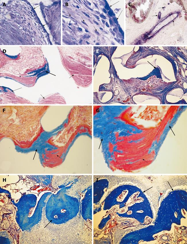
Figure 9 Intrinsic induction of bone formation by macroporous coral-derived hydroxyapatite matrices implanted in the rectus abdominis muscle of non-human primates of the species P.
ursinus. A-C: Morphology of cellular differentiation and vascular invasion by coral-derived hydroxyapatite constructs implanted in the rectus abdominis muscle. Differentiation of osteoblastic-like cells at the hydroxyapatite interface with hyper chromatic nuclei facing the highly vascularized stroma (long arrows); short arrows in B indicate a stream of locomoting perivascular cells leading to the hydroxyapatite surface for further differentiation and morphogenesis; C: Capillaries penetrating the macroporous spaces are osteogenetic in Trueta definition[9] having endothelial/perivascular cells intensely alkaline phosphatase positive cells; the osteogenetic vessel induces further capillary sprouting and invasion (short arrows) also highly positive for alkaline phosphatase; D: Undecalcified section showing mineralized cellular condensations in blue facing the substratum (thick and thin long arrows), short arrows point to collagenous condensations as yet to be mineralized; E: Using particulated granular coral-derived macroporous constructs, the morphogenesis of bone is only found within a concavity of the implanted biomimetic matrix, short arrow points to vascular invasion and angiogenesis, long arrow points to newly formed bone within the concavity. D and E were instrumental to the realization that the concavity is the geometric shape that induces the ripple-like cascade of bone differentiation by induction within the implanted macroporous constructs; F, G: Mineralization (thick long arrows) of mesenchymal collagenic condensations (short arrows) at the interface of the macroporous construct; H, I: Generation of substantial bone formation by induction in macroporous coral-derived biomatrices when implanted in the rectus abdominis and harvested on day 90. Arrows point to newly formed bone in blue within the concavities of the biomimetic matrices. Undecalcified and decalcified sections cut at 6 μm and stained with Goldner’s trichrome and toluidine blue.
- Citation: Ripamonti U. Soluble and insoluble signals sculpt osteogenesis in angiogenesis. World J Biol Chem 2010; 1(5): 109-132
- URL: https://www.wjgnet.com/1949-8454/full/v1/i5/109.htm
- DOI: https://dx.doi.org/10.4331/wjbc.v1.i5.109


