Copyright
©The Author(s) 2016.
World J Biol Chem. Feb 26, 2016; 7(1): 110-127
Published online Feb 26, 2016. doi: 10.4331/wjbc.v7.i1.110
Published online Feb 26, 2016. doi: 10.4331/wjbc.v7.i1.110
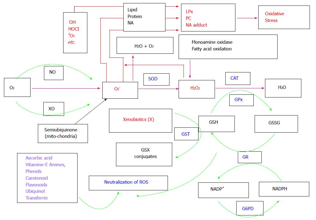
Figure 1 Pathways of active oxygen species metabolism.
OS physiology starts with O2 consumption in animals. O2 is incompletely reduced to super oxide radicals (O2-) via NO or XO or in mitochondria due incomplete reduction. O2- is dismutated to H2O2 by enzyme SOD. H2O2 is then scavenged by either CAT, or glutathione peroxidase with the help of the reduced glutathione (GSH, which form oxidised glutathione, i.e., GSSG). GSSG is recycled to GSH by the enzyme GR with the help of NADPH (which is converted into NADP+). NADP+ is reduced back to NADPH by the enzyme G6PD. GST is also responsible to remove xenobiotics (which are responsible to produce ROS) from cells with the help of GSH. Peroxiredoxins and thioredoxins system and glutaredoxins are responsible for scavenging H2O2 and reduction of other proteins (not shown in Figure). Redox regulatory non-enzymatic molecules such as ascorbic acid, flavonoids and phenols can also remove ROS such as OH, HOCL, O2, O2- and H2O2. With insufficient antioxidant defence, more ROS accumulation in cells occurs and it leads to oxidation of biomolecules such as proteins, lipids and nucleic acids to form PC, LPx and NA adducts, respectively. Formation of LPx, PC and NA leads to a disorder condition called as oxidative stress. OS: Oxidative stress; NO: Nitric oxidase; XO: Xanthine oxidase; SOD: Superoxide dismutase; CAT: Catalase; GR: Glutathione reductase; G6PD: Glucose-6-phosphate dehydrogenase; GST: Glutathione-S-transferase; PC: Protein carbonyls; LPx: Lipid peroxides; NA: Nucleic acid.
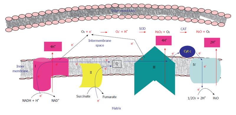
Figure 2 Production of reactive oxygen species in mitochondria.
ETC enzymes such as I (complex I), II (complex II), III (complex III) and IV (complex IV) are shown to be located in the inner membrane of mitochondria. During electron transport in ETC, e-via complex I (from NADH) and via complex II (from FADH2) are passed to complex III through Q and then subsequently passed to complex IV. In the sequence, they are finally delivered to the reaction in which O2 is reduced to H2O. In coupled with the above process, protons are pumped to intermembrane space via complexes I, III and IV. This forms a chemiosmotic gradient to avail free energy for synthesis of ATP molecules via complex V enzyme (not shown in the figure). However, due to leakage of electrons at complex I and complex III enzymes during electron transport, O2 molecules are incompletely reduced to form ROS such as O2- anion radical, H2O2 and OH radical (formed due to the interaction between O2- and H2O2). Enzymes such as SOD and CAT catalyzes the reactions at the respective steps as shown in the figure. It leads to production of subsequent other ROS. Blue thick arrows indicate direction of flow of electrons in ETC. Red arrows indicate pumping of protons from matrix to intermembrane space. Black arrows indicate leakage of electrons to intermembrane space[40]. ETC: Electron transport chain; e-: Electrons; Q: Ubiquinol; O2-: Superoxide; H2O2: Hydrogen peroxide; OH: Hydrogen; SOD: Superoxide dismutase; CAT: Catalase.
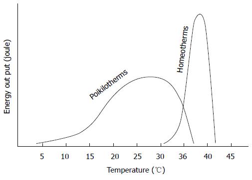
Figure 3 A proposed energy production system under rise in temperature condition in poikilotherms vs homeotherms.
Schematic comparison showing a sustained energy (joule) output between a poikilotherm and homeotherm as a function of core body temperature. The homeotherms have a much higher energy output range but can only function over a very narrow range of body temperatures (30 °C-40 °C in rats). However, poikilotherms have a much lower energy output range but can function over a very wide range of body temperatures (5 °C-40 °C in house lizards). Since, the energy output range is different in different animals, values are not provided in Y-axis[159].
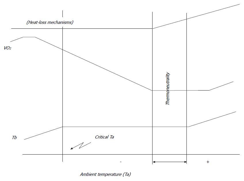
Figure 4 Relation among O2 consumption increases in ambient temperature and thermoregulation in animals.
When ambient Ta decreases below thermoneutrality, VO2 increases maintaining body Tb. When thermogenesis does not suffice, Tb begins to fall (critical Ta). The ability to maintain a thermoneutral range is mostly due to an increase in heat dissipation. Eventually, with further increases in Ta, heat-loss mechanisms will not prevent a rise in Tb, which will also lead to a rise in VO2[162]. Ta: Temperature; VO2: Oxygen consumption; Tb: Temperature.
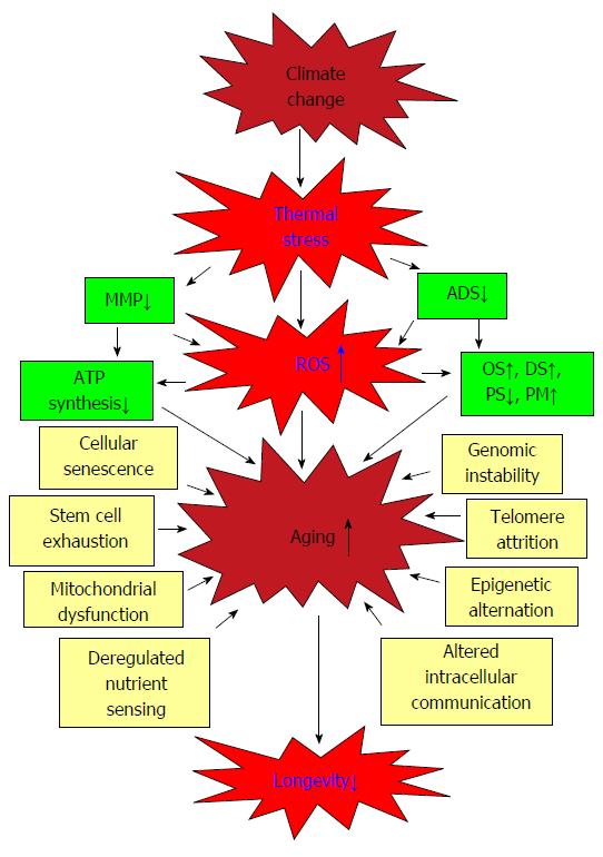
Figure 5 Possible correlation between global warming and longevity modulated by oxidative metabolism in animals.
Climate change is responsible to increase the mean global temperature which may lead to produce ROS in susceptible animals. Increase IT can also be responsible to produce ROS via diminishing ADS and MMP. This in turn may increase aging in animals by elevating oxidative stress. Other vital processes such as decrease in ATP synthesis, increasing the chance to DS, PS and PM in animals can lead to additional ROS production and thereby, shortening their longevity. Due to thermal stress, physiological processes such as genomic instability, telomere shortening and mitochondrial dysfunction can also lead to elevate aging in animals which may ultimately decrease their longevity. ROS: Reactive oxygen species; IT: In temperature; ADS: Antioxidant defence system; MMP: Mitochondrial membrane potential; DS: Disease susceptibility; PS: Proteostasis; PM: Protein misfolding.
- Citation: Paital B, Panda SK, Hati AK, Mohanty B, Mohapatra MK, Kanungo S, Chainy GBN. Longevity of animals under reactive oxygen species stress and disease susceptibility due to global warming. World J Biol Chem 2016; 7(1): 110-127
- URL: https://www.wjgnet.com/1949-8454/full/v7/i1/110.htm
- DOI: https://dx.doi.org/10.4331/wjbc.v7.i1.110


