Copyright
©2013 Baishideng Publishing Group Co.
World J Gastrointest Surg. Mar 27, 2013; 5(3): 51-61
Published online Mar 27, 2013. doi: 10.4240/wjgs.v5.i3.51
Published online Mar 27, 2013. doi: 10.4240/wjgs.v5.i3.51
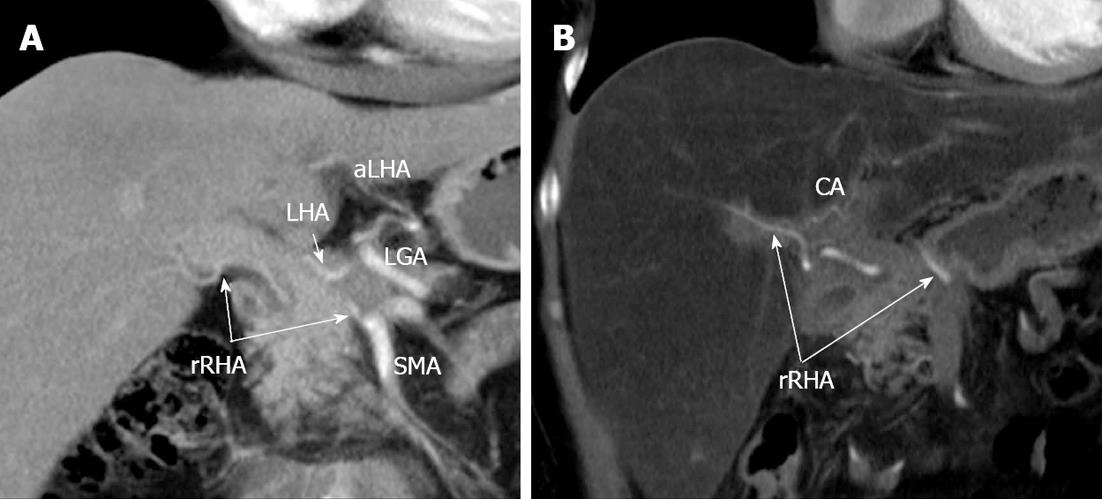
Figure 12 Coronal image.
Arterial phase. A: Previous to surgery. Aberrant arterial vasculature (Michels, type VIIIb): replaced right hepatic artery (rRHA) stemming from superior mesenteric artery (SMA), the left gastric artery (LGA) giving rise to accessorial left hepatic artery (aLHA). No interlobar collateral is detectable; B: Distal pancreatectomy with excision of celiac artery (CA), LGA, common hepatic (CHA) and left hepatic (LHA) arteries. Increased blood flow via rRHA is displayed and extraparenchymal hilar interlobar collateral transmitting blood supply to left hepatic lobe became visible.
- Citation: Egorov VI, Petrov RV, Lozhkin MV, Maynovskaya OA, Starostina NS, Chernaya NR, Filippova EM. Liver blood supply after a modified Appleby procedure in classical and aberrant arterial anatomy. World J Gastrointest Surg 2013; 5(3): 51-61
- URL: https://www.wjgnet.com/1948-9366/full/v5/i3/51.htm
- DOI: https://dx.doi.org/10.4240/wjgs.v5.i3.51


