Copyright
©2013 Baishideng Publishing Group Co.
World J Gastrointest Surg. Mar 27, 2013; 5(3): 51-61
Published online Mar 27, 2013. doi: 10.4240/wjgs.v5.i3.51
Published online Mar 27, 2013. doi: 10.4240/wjgs.v5.i3.51
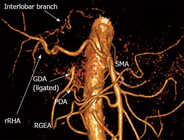
Figure 11 Three-dimensional computed tomography angiography subsequent to distal pancreatectomy with excision of celiac artery, left gastric, common hepatic and left hepatic arteries.
Blood supply to right hepatic lobe is provided by superior mesenteric artery (SMA) through the replaced right hepatic artery (rRHA) and that to left hepatic lobe - via interlobar collateral anastomosing with rRHA. Stomach is supplied from SMA via pancreaticoduodenal artery (PDA) and, thereafter, through gastroduodenal artery (GDA) and right gastro-epiploic artery (RGEA).
- Citation: Egorov VI, Petrov RV, Lozhkin MV, Maynovskaya OA, Starostina NS, Chernaya NR, Filippova EM. Liver blood supply after a modified Appleby procedure in classical and aberrant arterial anatomy. World J Gastrointest Surg 2013; 5(3): 51-61
- URL: https://www.wjgnet.com/1948-9366/full/v5/i3/51.htm
- DOI: https://dx.doi.org/10.4240/wjgs.v5.i3.51


