Copyright
©2013 Baishideng Publishing Group Co.
World J Gastrointest Surg. Mar 27, 2013; 5(3): 51-61
Published online Mar 27, 2013. doi: 10.4240/wjgs.v5.i3.51
Published online Mar 27, 2013. doi: 10.4240/wjgs.v5.i3.51
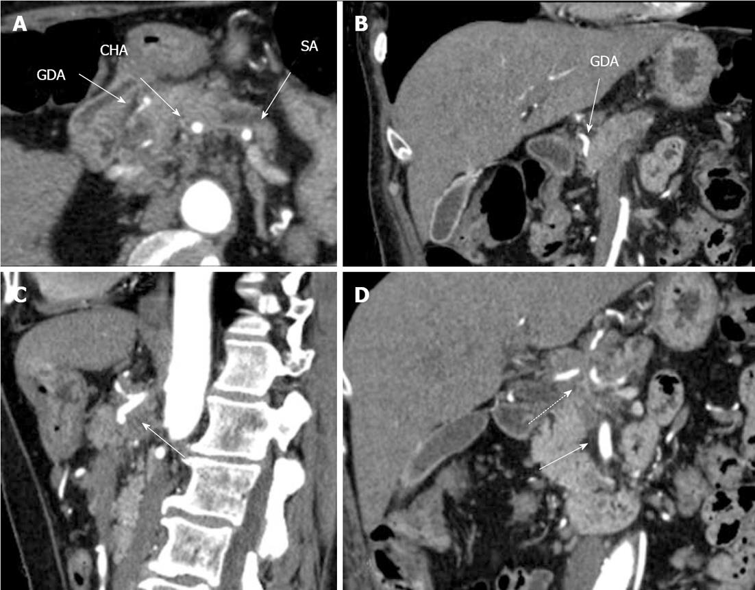
Figure 1 Preoperative computed tomography.
Arterial phase. A: Axial image. The common hepatic (CHA) and splenic (SA) arteries present circumferential adjacency to pancreatic body ductal adenocarcinoma. The gastroduodenal artery (GDA) appears to be completely encircled by tumor; B: Frontal view. Computed tomography (CT) evidences circumferential infiltration of GDA; C: The celiac artery (CA) along with CHA springing from it, are completely circumscribed by tumor (arrow); D. All three CA branches (dashed arrow) show circumferential tumor contact. The superior mesenteric artery is unaffected (arrow).
- Citation: Egorov VI, Petrov RV, Lozhkin MV, Maynovskaya OA, Starostina NS, Chernaya NR, Filippova EM. Liver blood supply after a modified Appleby procedure in classical and aberrant arterial anatomy. World J Gastrointest Surg 2013; 5(3): 51-61
- URL: https://www.wjgnet.com/1948-9366/full/v5/i3/51.htm
- DOI: https://dx.doi.org/10.4240/wjgs.v5.i3.51


