Copyright
©2010 Baishideng Publishing Group Co.
World J Gastrointest Surg. Aug 27, 2010; 2(8): 270-274
Published online Aug 27, 2010. doi: 10.4240/wjgs.v2.i8.270
Published online Aug 27, 2010. doi: 10.4240/wjgs.v2.i8.270
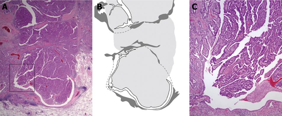
Figure 2 Pathological findings of resected lung tumor (HE staining).
The illustration (B) depicts the location and growth pattern of the tumor. A tumor arose from the bronchial wall and grew endoluminally. The tumor, forming a polypoid mass, almost completely obstructed the middle lobe bronchus. The tumor measured approximately 3 cm in diameter. The extent of the tumor is illustrated in light gray. Dark gray areas indicate smooth muscle layers of the bronchial wall. Asterisks indicate the remaining original bronchial lumen. Histologically, the tumor was a well-differentiated papillary and tubular adenocarcinoma. Figure 2C is a magnification of the part enclosed by a grid in Figure 2A. The tumor cells displaced the bronchial mucosal epithelium. Original magnification: ×10 (A), ×40 (C).
- Citation: Hanyu T, Kanda T, Matsuki A, Hasegawa G, Yajima K, Tsuchida M, Kosugi SI, Naito M, Hatakeyama K. Endobronchial metastasis from adenocarcinoma of gastric cardia 7 years after potentially curable resection. World J Gastrointest Surg 2010; 2(8): 270-274
- URL: https://www.wjgnet.com/1948-9366/full/v2/i8/270.htm
- DOI: https://dx.doi.org/10.4240/wjgs.v2.i8.270


