
















©The Author(s) 2025.
World J Gastrointest Surg. Aug 27, 2025; 17(8): 109213
Published online Aug 27, 2025. doi: 10.4240/wjgs.v17.i8.109213
Published online Aug 27, 2025. doi: 10.4240/wjgs.v17.i8.109213
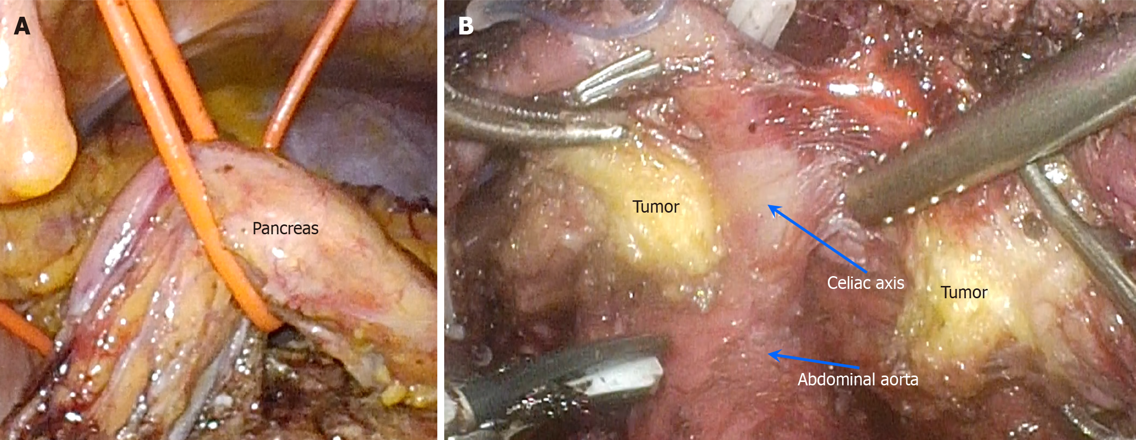
Figure 3 Intraoperative images.
A: A 10-Fr red rubber urinary catheter was passed beneath the pancreas and suspended from the abdominal wall, effectively exposing the retroperitoneal space; B: An ultrasonic scalpel was used to incise the tumor along the vascular axis, thereby exposing the celiac axis. The vascular wall remained intact without evidence of tumor cell infiltration or encroachment. Arrow: Correspondingly mark the blood vessels.
- Citation: Wu GZ, Fang SZ, Yu SA, Yu M. Resection of a ganglioneuroma encasing major blood vessels using three-dimensional laparoscopy combined with organ suspension: A case report. World J Gastrointest Surg 2025; 17(8): 109213
- URL: https://www.wjgnet.com/1948-9366/full/v17/i8/109213.htm
- DOI: https://dx.doi.org/10.4240/wjgs.v17.i8.109213