
















©The Author(s) 2025.
World J Gastrointest Surg. Aug 27, 2025; 17(8): 109213
Published online Aug 27, 2025. doi: 10.4240/wjgs.v17.i8.109213
Published online Aug 27, 2025. doi: 10.4240/wjgs.v17.i8.109213
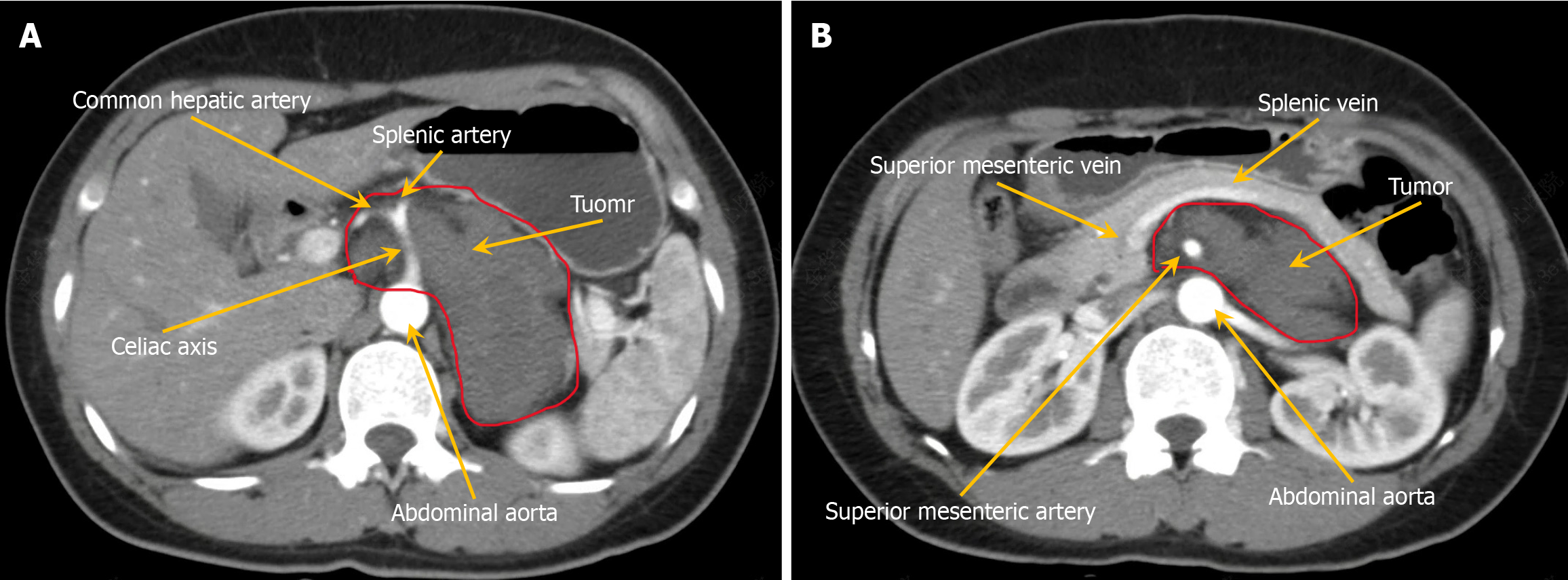
Figure 1 Enhanced computed tomography scan depicting a tumor located posterior to the pancreas and adjacent to the hepatic portal vein with progressive enhancement.
The maximum cross-sectional dimensions of the tumor are approximately 8.5 cm × 5 cm. A: The tumor completely encases the celiac axis, common hepatic artery, and splenic artery and is closely adjacent to the abdominal aorta; however, no notable luminal stenosis is observed in these vessels; B: The tumor completely encases the superior mesenteric artery and is closely associated with the superior mesenteric, splenic, and left renal veins. Arrow: Correspondingly mark the blood vessels or tumors; Red line: The boundary of the tumor.
- Citation: Wu GZ, Fang SZ, Yu SA, Yu M. Resection of a ganglioneuroma encasing major blood vessels using three-dimensional laparoscopy combined with organ suspension: A case report. World J Gastrointest Surg 2025; 17(8): 109213
- URL: https://www.wjgnet.com/1948-9366/full/v17/i8/109213.htm
- DOI: https://dx.doi.org/10.4240/wjgs.v17.i8.109213