Copyright
©The Author(s) 2025.
World J Gastrointest Surg. Aug 27, 2025; 17(8): 108959
Published online Aug 27, 2025. doi: 10.4240/wjgs.v17.i8.108959
Published online Aug 27, 2025. doi: 10.4240/wjgs.v17.i8.108959
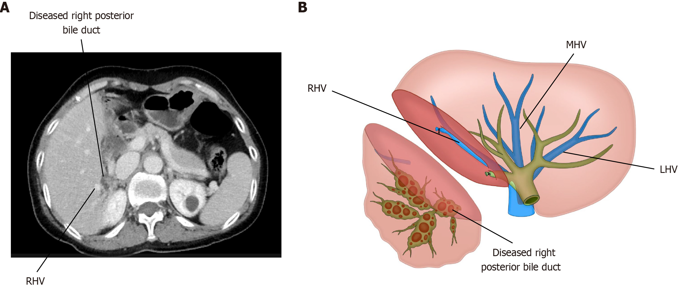
Figure 5 Diagram of the lesion bile duct.
A: Dilated bile ducts and stones, which are often close to the iconic hepatic veins; B: Hepatectomy was performed along the double markers of diseased bile duct and hepatic vein. MHV: Middle hepatic vein; LHV: Left hepatic vein; RHV: Right hepatic vein.
- Citation: Yang YH, Li XJ, Liu YX, Wang XR, Li JW. Laparoscopic hepatectomy based on diseased bile duct tree territory guided by double landmarks for hepatolithiasis: A case report. World J Gastrointest Surg 2025; 17(8): 108959
- URL: https://www.wjgnet.com/1948-9366/full/v17/i8/108959.htm
- DOI: https://dx.doi.org/10.4240/wjgs.v17.i8.108959


