
















©The Author(s) 2025.
World J Gastrointest Surg. Aug 27, 2025; 17(8): 108959
Published online Aug 27, 2025. doi: 10.4240/wjgs.v17.i8.108959
Published online Aug 27, 2025. doi: 10.4240/wjgs.v17.i8.108959
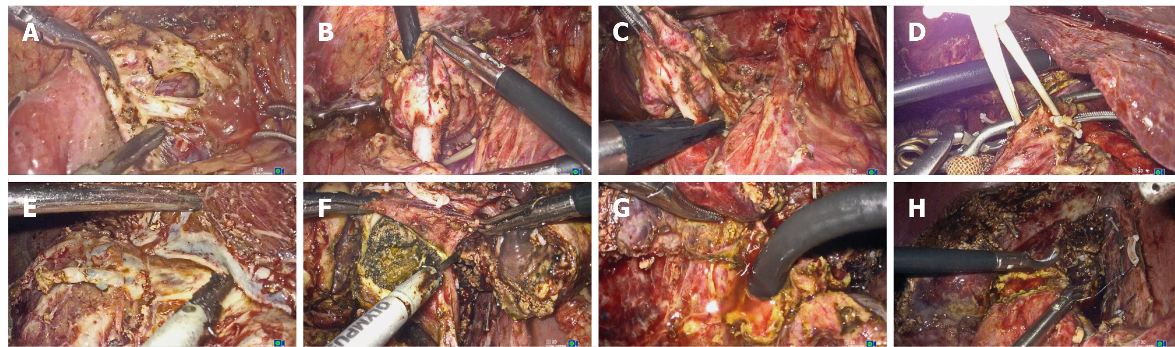
Figure 3 Intraoperative images during the operation.
A: Adhesiolysis and mobilization of the extrahepatic bile duct; B: Cystic dilatation of the extrahepatic bile duct was identified. Choledochotomy was performed, followed by choledochoscopic exploration, stone extraction, and assessment of the duodenal papilla; C: Resection of the dilated bile duct; D: Intermittent occlusion of the right anterior pedicle; E: Hepatic transection guided by the pathological bile ducts and hepatic veins; F: Opening of the hilar bile ducts with complete stone clearance; G: Repeat choledochoscopic exploration of the right anterior sectoral bile ducts; H: Hepa
- Citation: Yang YH, Li XJ, Liu YX, Wang XR, Li JW. Laparoscopic hepatectomy based on diseased bile duct tree territory guided by double landmarks for hepatolithiasis: A case report. World J Gastrointest Surg 2025; 17(8): 108959
- URL: https://www.wjgnet.com/1948-9366/full/v17/i8/108959.htm
- DOI: https://dx.doi.org/10.4240/wjgs.v17.i8.108959