Copyright
©The Author(s) 2025.
World J Gastrointest Surg. Aug 27, 2025; 17(8): 108579
Published online Aug 27, 2025. doi: 10.4240/wjgs.v17.i8.108579
Published online Aug 27, 2025. doi: 10.4240/wjgs.v17.i8.108579
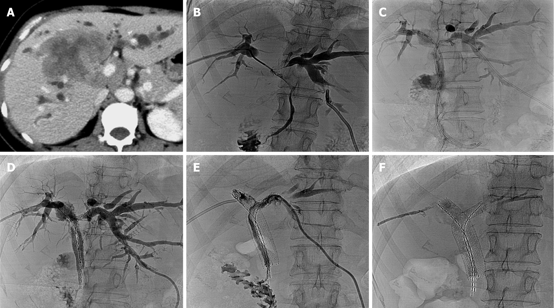
Figure 3 Hilar cholangiocarcinoma in a 49-year-old female patient.
A: Abdominal contrast-enhanced computed tomography shows hilar cho
- Citation: Zhou CG, Zhang Y, Li H, Liu KY, Yang XY, Gao K. Outcomes of iodine-125 seed strips combined with double self-expandable metallic stent for Bismuth type III and IV malignant biliary obstruction. World J Gastrointest Surg 2025; 17(8): 108579
- URL: https://www.wjgnet.com/1948-9366/full/v17/i8/108579.htm
- DOI: https://dx.doi.org/10.4240/wjgs.v17.i8.108579


