Copyright
©The Author(s) 2025.
World J Gastrointest Surg. Jun 27, 2025; 17(6): 107866
Published online Jun 27, 2025. doi: 10.4240/wjgs.v17.i6.107866
Published online Jun 27, 2025. doi: 10.4240/wjgs.v17.i6.107866
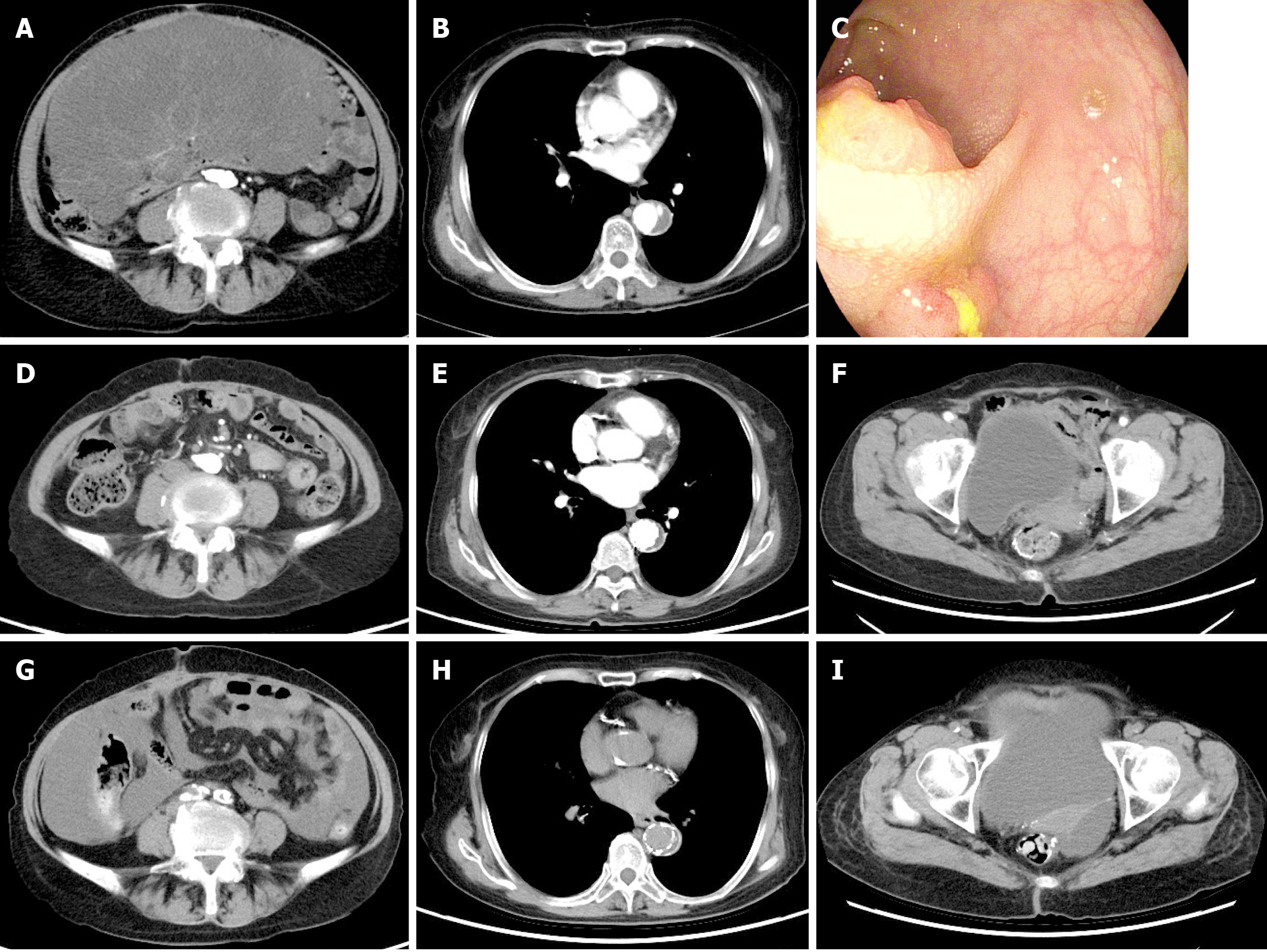
Figure 1 Preoperative ancillary investigations following hospital admission and ancillary investigations before discharge.
A: Contrast-enhanced abdominal computed tomography (CT) showing a large intra-abdominal tumor measuring approximately 25 cm × 20 cm; B: Contrast-enhanced chest CT revealing an eccentric, curvilinear filling defect in the thoracic aorta, accompanied by mild luminal stenosis and rigidity; C: Electronic colonoscopy showing a small protruding lesion in the rectum occupying the lumen for about 1/4 weeks, with central ulceration, and a stiff, brittle texture; D-F: First postoperative thoracic and abdominal CT review; G-I: Second postoperative thoracic and abdominal CT review. Comparative analysis across the three image sets demonstrates the progression and response to treatment.
- Citation: Wang M, Sun J, Song ZQ, Chen XQ, Xie GD, Zhu Y, Zhou YK. Giant transverse colonic mesenteric mucinous liposarcoma combined with rectal cancer and aortic coarctation: A case report and review of literature. World J Gastrointest Surg 2025; 17(6): 107866
- URL: https://www.wjgnet.com/1948-9366/full/v17/i6/107866.htm
- DOI: https://dx.doi.org/10.4240/wjgs.v17.i6.107866


