Copyright
©The Author(s) 2025.
World J Gastrointest Surg. Jun 27, 2025; 17(6): 107351
Published online Jun 27, 2025. doi: 10.4240/wjgs.v17.i6.107351
Published online Jun 27, 2025. doi: 10.4240/wjgs.v17.i6.107351
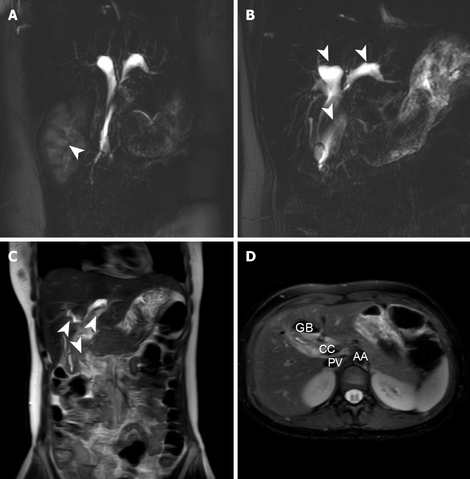
Figure 4 Abdominal magnetic resonance imaging findings at second admission.
A: Resolution of hydronephrosis (white arrow); B: Persistent dilatation of intra- and extrahepatic biliary ducts (white arrow); C: There was significant reduction in abdominal cyst size (white arrow); D: Key anatomical structures of the abdomen. GB: Gall bladder; CC: Choledochal cyst; PV: Portal vein; AA: Abdominal aorta.
- Citation: Wang DD, Du YY, Li YZ, Wang W, Ma TL, Xu XC, Mi C, Wang SY, Cui F, She YH, Wang MC, Yang HT. Treatment of giant choledochal cysts with combined surgery and percutaneous transhepatic cholangial drainage: A case report. World J Gastrointest Surg 2025; 17(6): 107351
- URL: https://www.wjgnet.com/1948-9366/full/v17/i6/107351.htm
- DOI: https://dx.doi.org/10.4240/wjgs.v17.i6.107351


