Copyright
©The Author(s) 2025.
World J Gastrointest Surg. Jun 27, 2025; 17(6): 107235
Published online Jun 27, 2025. doi: 10.4240/wjgs.v17.i6.107235
Published online Jun 27, 2025. doi: 10.4240/wjgs.v17.i6.107235
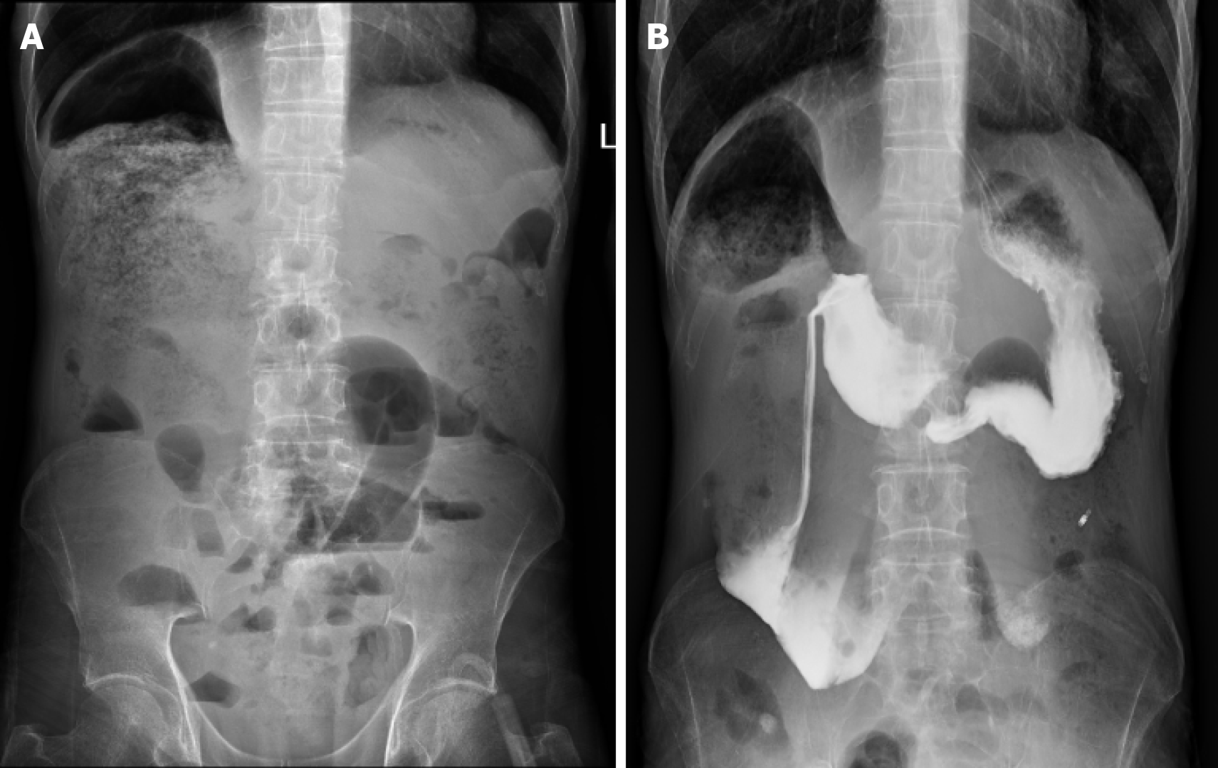
Figure 1 Abdominal imaging findings.
A: Upright abdominal radiograph showing an elevated right diaphragm, with dilated intestinal loops below the diaphragm containing significant content. Small bowel distension in the lower abdomen with multiple gas-fluid levels are also visible; B: Upper gastrointestinal contrast study showing marked dilatation of the duodenal bulb, and descending and horizontal segments, with the widest area measuring approximately 6 cm. No duodenal folds are observed.
- Citation: Jiang S, Zhou YX, Sun XH, Chen PP, Tang H, Chen Y, Liu YP, Li YX, Kang L. Hereditary chronic intestinal pseudo-obstruction caused by a rare MYH11 mutation: A case report. World J Gastrointest Surg 2025; 17(6): 107235
- URL: https://www.wjgnet.com/1948-9366/full/v17/i6/107235.htm
- DOI: https://dx.doi.org/10.4240/wjgs.v17.i6.107235


