Copyright
©The Author(s) 2024.
World J Gastrointest Surg. Mar 27, 2024; 16(3): 966-973
Published online Mar 27, 2024. doi: 10.4240/wjgs.v16.i3.966
Published online Mar 27, 2024. doi: 10.4240/wjgs.v16.i3.966
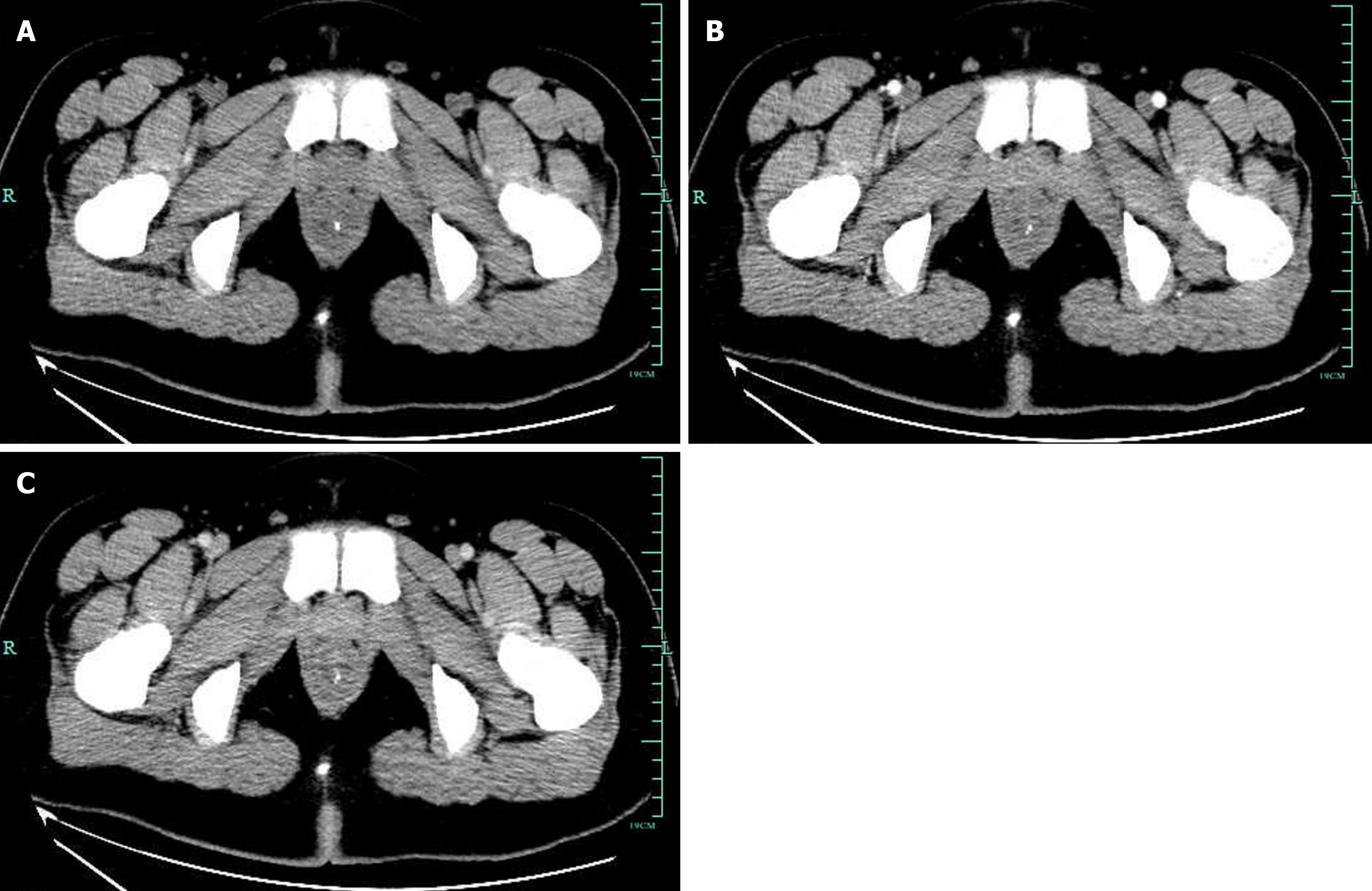
Figure 1 Enhanced total abdominal computed tomography showing that part of the rectum was thickened with edema and numerous tiny calcification foci indicative of phleboliths.
A: Plain scan; B: Enhancement scan; C: Venous scan.
- Citation: Zhu HT, Chen WG, Wang JJ, Guo JN, Zhang FM, Xu GQ, Chen HT. Endoscopic ultrasound-guided lauromacrogol injection for treatment of colorectal cavernous hemangioma: Two case reports. World J Gastrointest Surg 2024; 16(3): 966-973
- URL: https://www.wjgnet.com/1948-9366/full/v16/i3/966.htm
- DOI: https://dx.doi.org/10.4240/wjgs.v16.i3.966


