Copyright
©The Author(s) 2022.
World J Gastrointest Surg. Oct 27, 2022; 14(10): 1131-1140
Published online Oct 27, 2022. doi: 10.4240/wjgs.v14.i10.1131
Published online Oct 27, 2022. doi: 10.4240/wjgs.v14.i10.1131
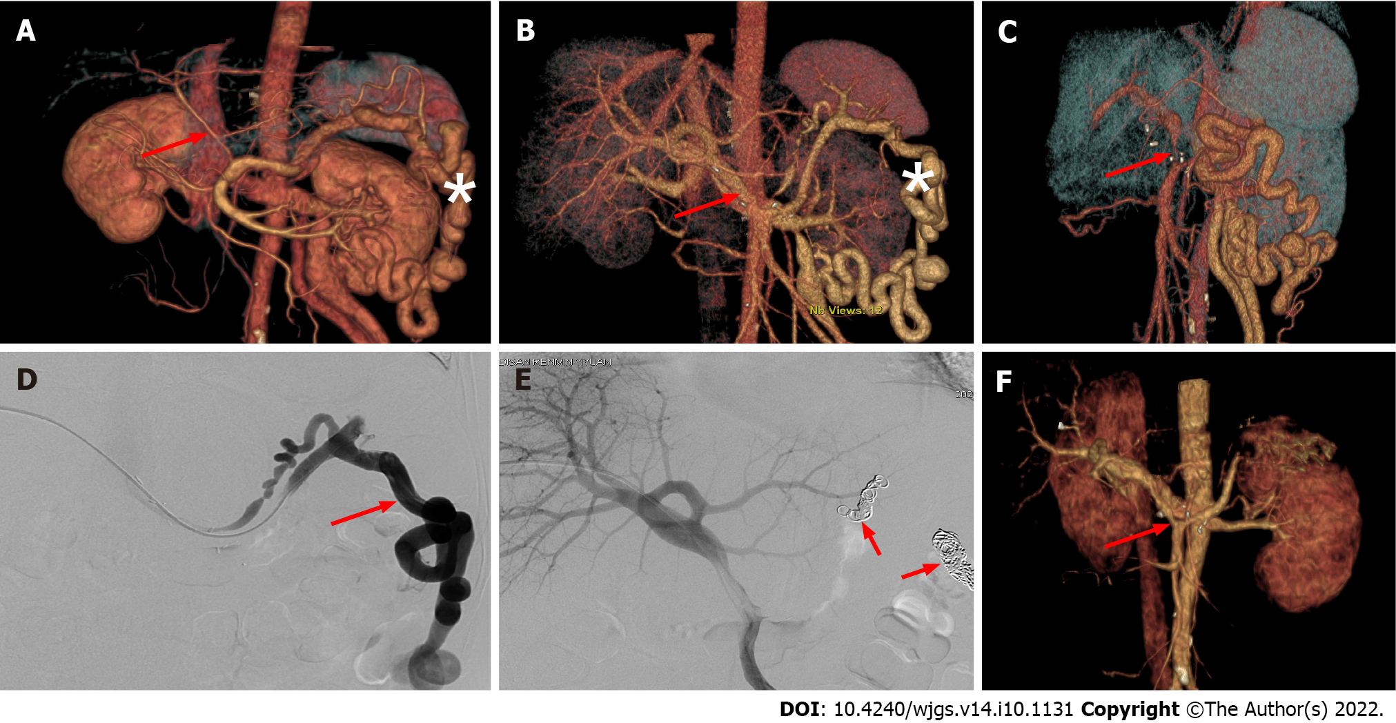
Figure 2 Embolization of large splenorenal shunt under digital subtraction angiography alleviates portal vein insufficiency after liver transplantation.
A: Preoperative three-dimensional (3D) visualization model showed a slender portal vein (arrow) and obvious splenorenal shunt varices (*); B: Postoperative 3D visualization model on day 3 showed a normal portal vein shape and unobstructed blood flow (arrow), and splenorenal shunt varicosity was reduced (*); C: Postoperative 3D visualization model (at 3 wk) showed portal vein stenosis in the initial segment (arrow), and color Doppler ultrasound examination indicated insufficient portal venous blood supply; D: Percutaneous and transhepatic splenic venography showed that most splenic venous flow drained into the inferior vena cava through the splenorenal shunt, but did not drain into the portal vein (arrow); E: After embolization of the splenorenal shunt (arrow), angiography showed that blood flow was mainly present into the portal vein; F: 3D visualization model 1 wk after the vascular intervention showed unobstructed portal vein flow (arrow), and the splenorenal shunt was no longer visible.
- Citation: Zhao D, Huang YM, Liang ZM, Zhang KJ, Fang TS, Yan X, Jin X, Zhang Y, Tang JX, Xie LJ, Zeng XC. Reconstructing the portal vein through a posterior pancreatic tunnel: New choice for portal vein thrombosis during liver transplantation. World J Gastrointest Surg 2022; 14(10): 1131-1140
- URL: https://www.wjgnet.com/1948-9366/full/v14/i10/1131.htm
- DOI: https://dx.doi.org/10.4240/wjgs.v14.i10.1131


