Copyright
©The Author(s) 2019.
World J Gastrointestinal Surgery. Apr 27, 2019; 11(4): 229-236
Published online Apr 27, 2019. doi: 10.4240/wjgs.v11.i4.229
Published online Apr 27, 2019. doi: 10.4240/wjgs.v11.i4.229
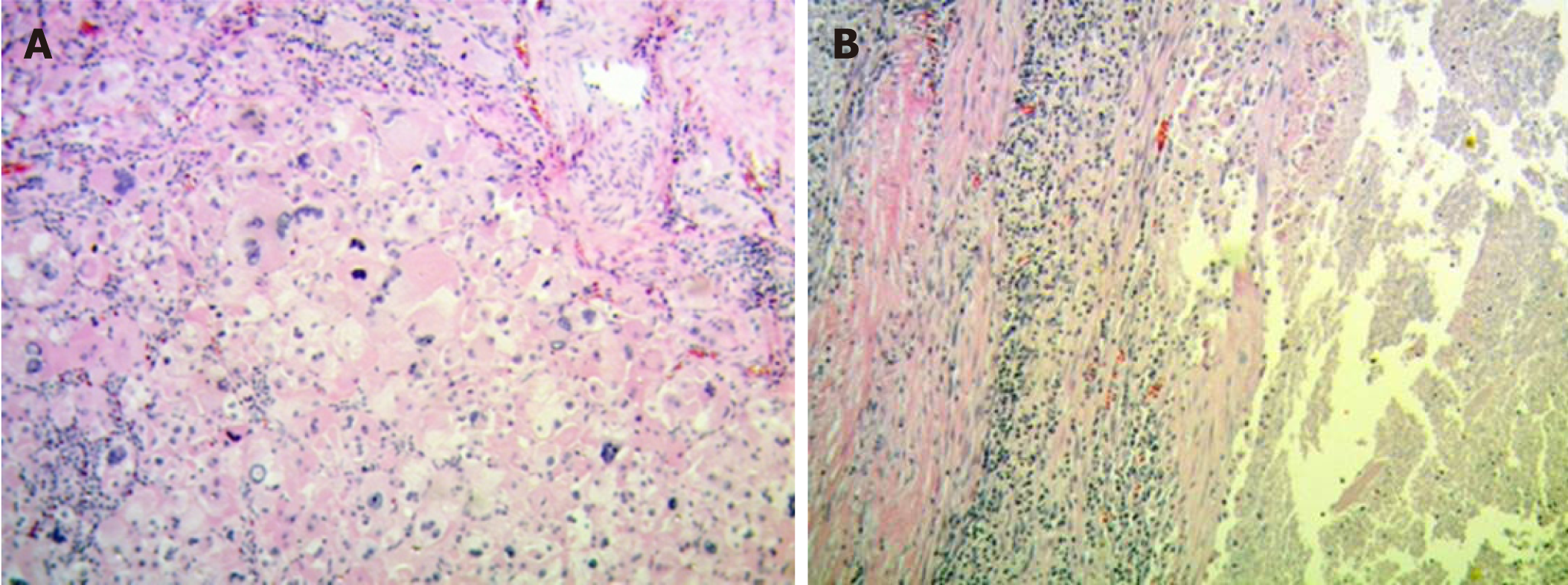
Figure 4 Hematoxilin and eosin.
A: With 400×, tumor area with atypia and abundant mitotic figures can be noticed; B: With 100×, sector with extensive necrosis and the presence of a tumor capsule can be seen.
- Citation: Glinka J, Clariá RS, Fratanoni E, Spina J, Mullen E, Ardiles V, Mazza O, Pekolj J, de Santibañes M, de Santibañes E. Malignant transformation of hepatocellular adenoma in a young female patient after ovulation induction fertility treatment: A case report. World J Gastrointestinal Surgery 2019; 11(4): 229-236
- URL: https://www.wjgnet.com/1948-9366/full/v11/i4/229.htm
- DOI: https://dx.doi.org/10.4240/wjgs.v11.i4.229


