Copyright
©The Author(s) 2019.
World J Gastrointestinal Surgery. Apr 27, 2019; 11(4): 229-236
Published online Apr 27, 2019. doi: 10.4240/wjgs.v11.i4.229
Published online Apr 27, 2019. doi: 10.4240/wjgs.v11.i4.229
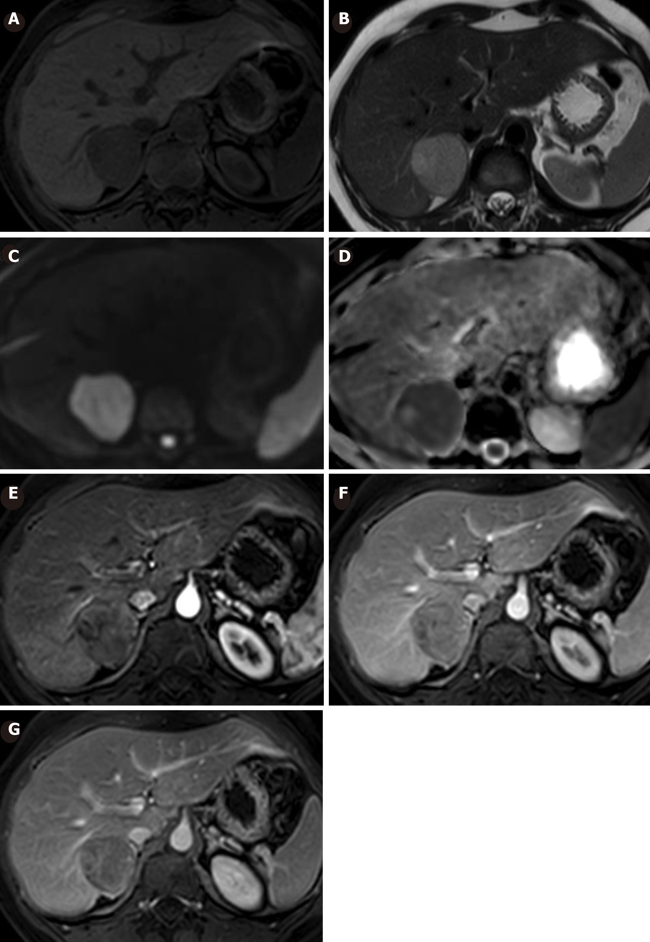
Figure 3 Magnetic resonance imaging showing enlargement of the lesion in follow-up.
A: It is hypointense in T1; B: Hyperintense in T2; C: Hyperintense in diffusion-weighted image (DWI) b800. D: It shows restriction in ADC; E: Has slight heterogeneous enhancement in arterial phase; F: Has more evident enhancement in portal phase; G: Also more enhancement in late phase. The increase in size and the presence of restriction in DWI is suggestive of malignant transformation.
- Citation: Glinka J, Clariá RS, Fratanoni E, Spina J, Mullen E, Ardiles V, Mazza O, Pekolj J, de Santibañes M, de Santibañes E. Malignant transformation of hepatocellular adenoma in a young female patient after ovulation induction fertility treatment: A case report. World J Gastrointestinal Surgery 2019; 11(4): 229-236
- URL: https://www.wjgnet.com/1948-9366/full/v11/i4/229.htm
- DOI: https://dx.doi.org/10.4240/wjgs.v11.i4.229


