Copyright
©The Author(s) 2018.
World J Gastrointest Surg. Dec 27, 2018; 10(9): 107-110
Published online Dec 27, 2018. doi: 10.4240/wjgs.v10.i9.107
Published online Dec 27, 2018. doi: 10.4240/wjgs.v10.i9.107
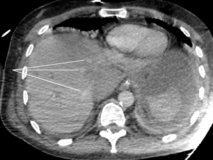
Figure 2 Injected abdominopelvic computed tomography showing extrinsic compression of the inferior vena cava and cavohepatic confluence.
Permeable hepatic veins are indicated by white lines.
- Citation: Dousse D, Bloom E, Suc B. Pancreaticoduodenectomy complicated by Budd-Chiari syndrome: A case report and review of literature. World J Gastrointest Surg 2018; 10(9): 107-110
- URL: https://www.wjgnet.com/1948-9366/full/v10/i9/107.htm
- DOI: https://dx.doi.org/10.4240/wjgs.v10.i9.107


