
















©The Author(s) 2015.
World J Diabetes. Mar 15, 2015; 6(2): 321-325
Published online Mar 15, 2015. doi: 10.4239/wjd.v6.i2.321
Published online Mar 15, 2015. doi: 10.4239/wjd.v6.i2.321
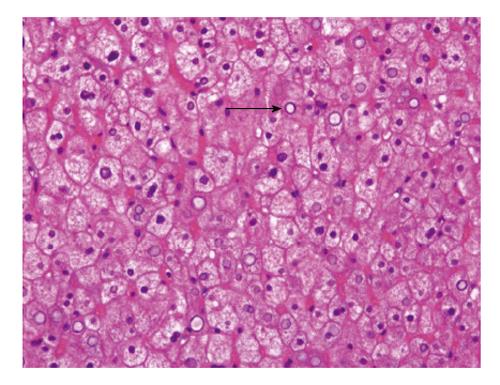
Figure 2 Liver biopsy, haematoxylin and eosin staining.
The hepatocytes are swollen with pale cytoplasm and accentuation of the cell membranes. Sinusoids appear compressed by the swollen hepatocytes. Glycogen nuclei are present (black arrow).
- Citation: Julián MT, Alonso N, Ojanguren I, Pizarro E, Ballestar E, Puig-Domingo M. Hepatic glycogenosis: An underdiagnosed complication of diabetes mellitus? World J Diabetes 2015; 6(2): 321-325
- URL: https://www.wjgnet.com/1948-9358/full/v6/i2/321.htm
- DOI: https://dx.doi.org/10.4239/wjd.v6.i2.321